








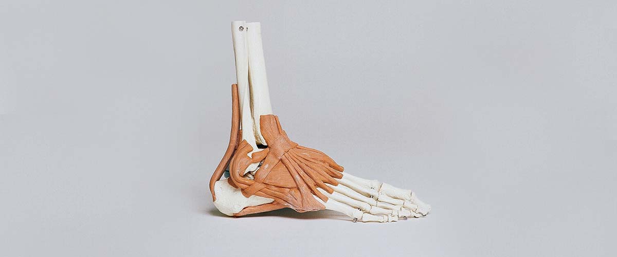
The posterior tibial tendon runs behind the bump on the inside of the ankle and supports the arch. When it becomes inflamed, degenerates or tears, the arch can gradually collapse. This is called posterior tibial tendon insufficiency or dysfunction (PTTI or PTTD), and it is the most important cause of adult-acquired flatfoot,1 now also called progressive collapsing foot deformity.2 It is common, especially in middle-aged and older adults.1,3
- Pain and swelling along the inside of the ankle and arch
- The arch flattens and the heel tilts outward over time
- Early stages: orthotics or a brace plus a structured exercise program; 83% succeeded in one study10
- Flexible flatfoot: tendon transfer and realignment osteotomies18,23
- Rigid or arthritic flatfoot: fusion of the hindfoot joints15,30
How it progresses
The condition is progressive and is usually described in stages:2,15
- Early: pain and swelling of the tendon with little or no change in foot shape.
- Flexible deformity: the arch flattens and the heel tilts out, but the foot can still be corrected by hand.
- Rigid deformity: the flatfoot becomes stiff, often with arthritis in the hindfoot joints.
- Ankle involvement: the deltoid ligament on the inside of the ankle fails and the ankle tilts, which is the hardest stage to correct.3
Degeneration within the tendon may make it more likely to tear under everyday loads.4
Symptoms
- Pain and swelling along the inside of the ankle and arch, worse with walking or standing
- A flattening arch, often noticed as one foot looking different from the other or shoes wearing unevenly
- Difficulty or pain rising up on the toes of one foot
- Later, pain on the outside of the ankle as the heel shifts and the bones pinch
Diagnosis
Dr. Dold checks the shape and flexibility of the foot, the heel position from behind, single-leg heel rise strength, and calf tightness (being unable to bring the ankle up about 10 degrees suggests a tight calf).14 Standing X-rays measure the arch and heel alignment. MRI or ultrasound evaluates the tendon and the spring ligament, and weight-bearing CT, when available, can help distinguish a stable flatfoot from a progressive collapsing deformity.6
Non-surgical treatment
Treatment usually starts without surgery,5,7 although high-quality studies of non-surgical options are limited.5
- Orthotics and bracing: medial heel-wedged inserts are effective in most early cases,8 and ankle-foot orthoses (such as an Arizona-type brace) have shown promising results for more advanced deformity.9
- A structured exercise program: with an orthosis and a structured program, 83% of 47 patients had successful outcomes and only 11% needed surgery.10 In a randomized trial, orthoses with eccentric exercise improved pain and function the most,11 and reviews support eccentric strengthening combined with stretching and orthoses.12,13
- Short-term immobilization in a walking boot for a painful flare
- Anti-inflammatory medicines and weight loss when appropriate7
Surgery for early disease
When the tendon stays painful despite good non-surgical care but the foot shape is still near normal, a tendoscopy (a small camera in the tendon sheath) can clean out the inflamed tissue. In one study, it relieved symptoms in 80% of cases,16 and combined with a heel-shift osteotomy it improved pain and alignment.17
Reconstruction of a flexible flatfoot
For a flexible flatfoot that still hurts, reconstruction combines procedures tailored to your foot:15,23
- Flexor digitorum longus (FDL) tendon transfer to replace the damaged posterior tibial tendon
- Medializing calcaneal osteotomy: shifting the heel bone inward. At long-term follow-up after tendon transfer and heel osteotomy, 87% were pain free and functioning well.18,19 A minimally invasive version has results comparable to open surgery.22
- Lateral column lengthening for a foot that points outward. In a randomized trial, it gave similar scores to the heel-shift osteotomy, corrected more, but had more joint degeneration.20,21
- Cotton osteotomy or first TMT fusion to support the inner column of the foot23
- Spring ligament repair when that ligament has failed26
- Calf lengthening (gastrocnemius recession) for a tight calf14
- Subtalar arthroereisis (a small spacer) is sometimes added, though sinus tarsi pain is a common complication.27,28
Patients improve significantly after reconstruction,23 walking speed and stride length improve,29 and patients over 65 improved as much as younger patients.24 Correcting the heel to a slightly inward (mild varus) position was associated with greater pain improvement.25
Rigid or arthritic flatfoot
When the flatfoot is stiff or arthritic, the hindfoot joints are realigned and fused. Triple arthrodesis gives predictable correction and pain relief, though arthritis in nearby joints can develop over time.32 In older adults, 14 of 17 were satisfied and all had less pain after triple arthrodesis.33 A medial double arthrodesis (fusing two joints through one incision) has shown similar benefit with fewer complications,30,31 and is often used today.
Recovery
- Non-surgical: most structured programs run several months.10
- After reconstruction or fusion: a period of non-weight-bearing in a splint and boot, followed by progressive weight-bearing and physical therapy. Full recovery takes many months.
Dr. Dold reviews your expected timeline based on your procedure and health.
Frequently asked questions
Is posterior tibial tendon dysfunction the same as flat feet?
It’s the most common cause of a flatfoot that develops in adulthood. Many people have lifelong flexible flat feet that never cause problems; PTTD is different because the arch collapses over time and is painful.
Will a brace or orthotic stop it from getting worse?
For many people with early or flexible disease, orthotics or a brace plus a structured exercise program relieve symptoms. In one study, 83% did well without surgery.
What happens if it’s not treated?
The deformity can progress and become rigid, sometimes with arthritis and, eventually, ankle involvement, which is harder to correct.
What does flatfoot reconstruction involve?
Usually a tendon transfer to replace the damaged tendon combined with bone cuts (osteotomies) to realign the heel and arch, plus calf lengthening or ligament repair when needed.
Am I too old for reconstruction?
Not necessarily. Patients over 65 improved as much as younger patients after flexible flatfoot reconstruction in one study.
Research cited on this page
- Smyth NA, Aiyer AA, Kaplan JR, et al. Adult-acquired flatfoot deformity. Eur J Orthop Surg Traumatol. 2017;27(4):433-439. PubMed
Loss of posterior tibial tendon function is the most important contributor to adult flatfoot, with an estimated prevalence over 3%. - Myerson MS, Thordarson DB, Johnson JE, et al. Classification and Nomenclature: Progressive Collapsing Foot Deformity. Foot Ankle Int. 2020;41(10):1271-1276. PubMed
An international consensus group proposed a new classification for progressive collapsing foot deformity to stage it and guide treatment. - Tiell JC, Malkamaki M, O’Connor P, et al. Chronic Deltoid Insufficiency in Stage IV Adult Acquired Flatfoot Deformity: Do We Have a Good Answer?. Cureus. 2024;16(6):e62711. PubMed
Adult flatfoot affects an estimated 10% of older adults, and stage IV deformity with deltoid failure remains hard to correct. - Mosier SM, Pomeroy G, Manoli A. Pathoanatomy and etiology of posterior tibial tendon dysfunction. Clin Orthop Relat Res. 1999(365):12-22. PubMed
The diseased tendon shows degenerative changes that may predispose it to rupture under normal loads. - Rhim HC, Dhawan R, Gureck AE, et al. Characteristics and Future Direction of Tibialis Posterior Tendinopathy Research: A Scoping Review. Medicina (Kaunas). 2022;58(12). PubMed
Initial treatment is typically non-surgical, but high-quality studies of non-surgical options are lacking. - Lalevée M, Dagneaux L, Lintz F, et al. Flatfoot: New diagnostic modalities. Orthop Traumatol Surg Res. 2026;112(1S):104415. PubMed
Weight-bearing CT helps distinguish a stable flatfoot from a progressive collapsing deformity. - Miniaci-Coxhead SL, Flemister AS. Office-based management of adult-acquired flatfoot deformity. Med Clin North Am. 2014;98(2):291-9. PubMed
The mainstays of non-surgical treatment are anti-inflammatories, weight loss and orthotic insoles or bracing. - Monteagudo M, Martínez-de-Albornoz P. Progressive Collapsing Foot Deformity. Is There Really a Johnson and Strom Stage I?. Foot Ankle Clin. 2021;26(3):443-463. PubMed
Medial heel-wedged orthoses are effective in most patients with early (stage I) disease. - Richie D. Biomechanics and Orthotic Treatment of the Adult Acquired Flatfoot. Clin Podiatr Med Surg. 2020;37(1):71-89. PubMed
Bracing with ankle-foot orthoses has shown promising results. - Alvarez RG, Marini A, Schmitt C, et al. Stage I and II posterior tibial tendon dysfunction treated by a structured nonoperative management protocol: an orthosis and exercise program. Foot Ankle Int. 2006;27(1):2-8. PubMed
With an orthosis and a structured exercise program, 83% of 47 patients had successful outcomes; 11% needed surgery. - Kulig K, Reischl SF, Pomrantz AB, et al. Nonsurgical management of posterior tibial tendon dysfunction with orthoses and resistive exercise: a randomized controlled trial. Phys Ther. 2009;89(1):26-37. PubMed
In a randomized trial, orthoses with eccentric exercise improved pain and function the most. - Ross MH, Smith MD, Mellor R, et al. Exercise for posterior tibial tendon dysfunction: a systematic review of randomised clinical trials and clinical guidelines. BMJ Open Sport Exerc Med. 2018;4(1):e000430. PubMed
Eccentric strengthening with stretching and orthoses had moderate effects on pain and disability. - Gómez-Jurado I, Juárez-Jiménez JM, Munuera-Martínez PV. Orthotic treatment for stage I and II posterior tibial tendon dysfunction (flat foot): A systematic review. Clin Rehabil. 2021;35(2):159-168. PubMed
Foot orthoses combined with exercise appeared to improve results in early-stage disease. - Chen L, Greisberg J. Achilles lengthening procedures. Foot Ankle Clin. 2009;14(4):627-37. PubMed
Being unable to bend the ankle up 10 degrees suggests a tight calf, which is common in adult flatfoot. - Henry JK, Shakked R, Ellis SJ. Adult-Acquired Flatfoot Deformity. Foot Ankle Orthop. 2019;4(1):2473011418820847. PubMed
Flexible deformities are usually treated with soft-tissue procedures and osteotomies; rigid deformities often need fusion. - Bernasconi A, Sadile F, Welck M, et al. Role of Tendoscopy in Treating Stage II Posterior Tibial Tendon Dysfunction. Foot Ankle Int. 2018;39(4):433-442. PubMed
Tendoscopy for stage II tendon dysfunction relieved symptoms in 80% of cases. - Saraiva D, Knupp M, Rodrigues AS, et al. Outcomes of Combined Posterior Tibial Tendon Tendoscopy and Medializing Calcaneal Osteotomy for Stage IA Progressive Collapsing Foot Deformity. Foot Ankle Int. 2023;44(7):629-636. PubMed
Tendoscopy combined with a heel-shift osteotomy for early deformity improved pain and alignment; most patients were very satisfied. - Chadwick C, Whitehouse SL, Saxby TS. Long-term follow-up of flexor digitorum longus transfer and calcaneal osteotomy for stage II posterior tibial tendon dysfunction. Bone Joint J. 2015;97-B(3):346-52. PubMed
At long-term follow-up after tendon transfer and heel osteotomy, 87% were pain free and functioning well. - Niki H, Hirano T, Okada H, et al. Outcome of medial displacement calcaneal osteotomy for correction of adult-acquired flatfoot. Foot Ankle Int. 2012;33(11):940-6. PubMed
After medial heel osteotomy with tendon transfer, scores improved from 59 to 91.3. - Osman AE, El-Gafary KA, Khalifa AA, et al. Medial displacement calcaneal osteotomy versus lateral column lengthening to treat stage II tibialis posterior tendon dysfunction, a prospective randomized controlled study. Foot (Edinb). 2021;47:101798. PubMed
In a randomized trial, heel-shift osteotomy and lateral column lengthening gave similar scores; lengthening corrected more but had more joint degeneration. - Modha RK, Kilmartin TE. Lateral Column Lengthening For Flexible Adult Acquired Flatfoot: Systematic Review and Meta-Analysis. J Foot Ankle Surg. 2021;60(6):1254-1269. PubMed
Lateral column lengthening gave excellent clinical and radiographic results; graft size under 8 mm mattered. - Vaggi S, Vitali F, Zanirato A, et al. Minimally invasive surgery in medial displacement calcaneal osteotomy for acquired flatfoot deformity: a systematic review of the literature. Arch Orthop Trauma Surg. 2024;144(3):1139-1147. PubMed
Minimally invasive heel osteotomy had about a 3% wound infection rate and results comparable to open surgery. - Conti MS, Garfinkel JH, Ellis SJ. Outcomes of Reconstruction of the Flexible Adult-acquired Flatfoot Deformity. Orthop Clin North Am. 2020;51(1):109-120. PubMed
Patients improve significantly after reconstruction of a flexible flatfoot tailored to their deformity. - Conti MS, Jones MT, Savenkov O, et al. Outcomes of Reconstruction of the Stage II Adult-Acquired Flatfoot Deformity in Older Patients. Foot Ankle Int. 2018;39(9):1019-1027. PubMed
Patients over 65 improved as much as younger patients after stage II reconstruction. - Conti MS, Ellis SJ, Chan JY, et al. Optimal Position of the Heel Following Reconstruction of the Stage II Adult-Acquired Flatfoot Deformity. Foot Ankle Int. 2015;36(8):919-27. PubMed
Correcting the heel to mild varus was associated with greater pain improvement. - García-Jarabo E, Ramos-Ramos LM, Sánchez-Morata EJ, et al. Surgical treatment at adult acquired flatfoot stage IIB: Spring ligament repair. Rev Esp Cir Ortop Traumatol. 2023;67(1):62-67. PubMed
Spring ligament repair with bony reconstruction improved scores from 52 to 88. - Baryeh KW, Ismail H, Sobti A, et al. Outcomes Following the Use of Subtalar Arthroereisis in the Correction of Adult Acquired Flatfoot: A Systematic Review. Foot Ankle Spec. 2022;15(4):384-393. PubMed
Subtalar arthroereisis as an adjunct improved scores with low complication rates, but higher-quality studies are needed. - Mattesi L, Ancelin D, Severyns MP. Is subtalar arthroereisis a good procedure in adult-acquired flatfoot? A systematic review of the literature. Orthop Traumatol Surg Res. 2021;107(6):103002. PubMed
Arthroereisis with other procedures gave good results, but complications (mainly sinus tarsi pain) were common. - Estes W, Syal A, Latt LD. Biomechanical Effects of Surgical Reconstruction for Flexible Progressive Collapsing Foot Deformity: A Systematic Review. Foot Ankle Spec. 2025;18(4):352-358. PubMed
Reconstruction of flexible deformity improved stride length, cadence and walking speed. - So E, Reb CW, Larson DR, et al. Medial Double Arthrodesis: Technique Guide and Tips. J Foot Ankle Surg. 2018;57(2):364-369. PubMed
Medial double arthrodesis has shown similar benefit to triple arthrodesis with fewer complications. - Bakaes Y, Gonzalez T, Hardin JW, et al. Comparison of the Acute Postoperative Complications Between Isolated Talonavicular Versus Talonavicular and Subtalar (Double) Arthrodesis in Flatfoot Deformity. Foot Ankle Spec. 2025;18(6):620-626. PubMed
Adding subtalar to talonavicular fusion did not raise 30-day complication risk. - Taylor R, Sammarco VJ. Minimizing the role of fusion in the rigid flatfoot. Foot Ankle Clin. 2012;17(2):337-49. PubMed
Triple arthrodesis gives predictable correction and pain relief, but adjacent arthritis can develop long-term. - Graves SC, Mann RA, Graves KO. Triple arthrodesis in older adults. Results after long-term follow-up. J Bone Joint Surg Am. 1993;75(3):355-62. PubMed
After triple arthrodesis in older adults, 14 of 17 were satisfied and all had less pain.
Medically reviewed by Andrew P. Dold, MD, FAOA, FAAOS, FACS, FRCSC. Last reviewed October 2026. This page is general information, not medical advice. Your own diagnosis and treatment plan depend on your exam and imaging.