









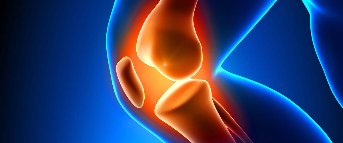
A plica is a fold of the knee’s lining (synovium) left over from development before birth. Almost everyone has one: a medial plica was present in 95% of knees in one arthroscopic study.6 Most never cause trouble, but after an injury or a rise in activity a plica can become thickened and inflamed and rub over the end of the thighbone. This is plica syndrome, a commonly overlooked cause of pain at the front of the knee.1,2
- Pain on the inner side of the kneecap, often with clicking or snapping when bending
- Most common in active teens and young adults1
- Ultrasound and a hands-on plica test are the most accurate diagnostic tools7,8
- Most patients improve with strengthening and stretching5,10
- Arthroscopic removal works well when non-surgical care fails1,11
Types of plica
- Medial patellar plica: the fold most often linked to symptoms, running along the inner side of the kneecap.1
- Suprapatellar plica: above the kneecap.22
- Infrapatellar plica: below the kneecap; should be considered a cause only after other problems are ruled out.20
- Lateral plica: uncommon, on the outer side.21
Symptoms
- Aching on the inner side of the kneecap, above the joint line3
- Clicking, snapping or catching when bending and straightening the knee
- Pain with stairs, squatting, running or sitting with the knee bent
- A tender, sometimes palpable band on the inside of the kneecap
Diagnosis
Plica syndrome can mimic other knee problems, so Dr. Dold examines the knee carefully and looks for other causes of anterior knee pain, such as patellofemoral pain or chondromalacia and meniscus tears.
- The medial patellar plica (MPP) test and ultrasound were the most accurate diagnostic tests, with sensitivity of about 90%; MRI was less accurate.7
- Dynamic ultrasound diagnosed symptomatic medial plica with 88% accuracy.8
- MRI reliably shows a plica and checks for other injuries, but is limited for classifying it.9
Because most people have a plica that causes no symptoms,6 finding one on MRI does not by itself mean it is the source of pain.
Non-surgical treatment
Treatment starts without surgery:3,4,10
- Relative rest and activity changes to calm the irritated plica
- Quadriceps strengthening and hamstring stretching: most patients respond to this program5
- Hip and lower-limb mechanics, footwear and training surfaces in runners23
- Anti-inflammatory medicines for a short course
- Cortisone injection into the plica or knee, which may help when given early1,5
In a pooled analysis of 527 patients, conservative and surgical treatment both improved function, supporting a conservative-first approach.10
Arthroscopic plica removal
If pain persists despite several months of good non-surgical care, the plica can be removed arthroscopically through small incisions. The procedure has low morbidity and results are generally good:1
- In young patients, pain fell from 7.9 to 3.1 on a 10-point scale with significantly better function.11
- Arthroscopic treatment was justified in young, very active athletes.13,14
- Results were most favorable when the plica was the only problem in the knee,15 with symptoms localized to the inner knee, onset after a rise in activity or a twist, and younger age.12
Why timing matters
A thick, inflamed plica can rub on and wear the cartilage of the inner femoral condyle and kneecap.16,18,19,24 Longer duration of symptoms was associated with cartilage damage, and patients with milder cases had better results after surgery.17 Chondromalacia predicted poorer results.12 If symptoms don’t improve with a dedicated program, it’s worth a re-evaluation rather than waiting indefinitely.
Recovery
- Non-surgical: most people improve over 6 to 12 weeks with a consistent strengthening and stretching program.
- After arthroscopy: walking the same day, physical therapy for strength and motion, and a return to sport usually within several weeks.
Dr. Dold reviews your expected timeline based on your treatment.
Frequently asked questions
Is a plica normal?
Yes. Most people have one, and it usually causes no problems. It becomes plica syndrome when it thickens and gets irritated after injury or overuse.
How do I know if my knee pain is from a plica?
Pain and clicking on the inner side of the kneecap, a tender band you can sometimes feel, and pain with bending are typical. A careful exam, and sometimes ultrasound or MRI, help rule out other causes.
Will plica syndrome go away on its own?
Many cases settle with rest, quadriceps strengthening and hamstring stretching. Some benefit from an injection, and a small number need arthroscopic removal.
Is plica surgery a big operation?
No. It’s an outpatient arthroscopy through small incisions, with walking the same day and therapy afterward.
Can a plica damage my knee?
A thick, inflamed plica can wear the cartilage it rubs against over time, which is one reason persistent symptoms should be evaluated.
Research cited on this page
- Schindler OS. ‘The Sneaky Plica’ revisited: morphology, pathophysiology and treatment of synovial plicae of the knee. Knee Surg Sports Traumatol Arthrosc. 2014;22(2):247-62. PubMed
The medial patellar plica is the fold most often linked to anteromedial knee pain; injections may help early, and arthroscopic excision gives good results in resistant cases. - Lee PYF, Nixion A, Chandratreya A, et al. Synovial Plica Syndrome of the Knee: A Commonly Overlooked Cause of Anterior Knee Pain. Surg J (N Y). 2017;3(1):e9-e16. PubMed
Synovial plica syndrome is a commonly overlooked cause of anterior knee pain, from injury or overuse. - Zmerly H, Akkawi I, Citarella R, et al. Clinical Management of Medial Patellar Plica Syndrome: Mini Review from Diagnosis to Treatment. Curr Rheumatol Rev. 2020;16(1):9-11. PubMed
Pain is typically just inside the kneecap above the joint line; treatment starts conservatively, with arthroscopic removal if that fails. - Sznajderman T, Smorgick Y, Lindner D, et al. Medial plica syndrome. Isr Med Assoc J. 2009;11(1):54-7. PubMed
Initial treatment includes physiotherapy, activity reduction and rest, then injection and anti-inflammatories if needed. - Griffith CJ, LaPrade RF. Medial plica irritation: diagnosis and treatment. Curr Rev Musculoskelet Med. 2008;1(1):53-60. PubMed
Most patients respond to quadriceps strengthening and hamstring stretching; a steroid injection may help those who do not. - Gurbuz H, Calpur OU, Ozcan M, et al. The synovial plicae in the knee joint. Saudi Med J. 2006;27(12):1839-42. PubMed
At arthroscopy, a medial (mediopatellar) plica was present in 95% of knees, so most plicae cause no symptoms. - Stubbings N, Smith T. Diagnostic test accuracy of clinical and radiological assessments for medial patella plica syndrome: a systematic review and meta-analysis. Knee. 2014;21(2):486-90. PubMed
The medial patellar plica test and ultrasound were the most accurate tests (sensitivity about 0.90); MRI was less accurate. - Paczesny L, Kruczynski J. Medial plica syndrome of the knee: diagnosis with dynamic sonography. Radiology. 2009;251(2):439-46. PubMed
Dynamic ultrasound diagnosed symptomatic medial plica with 88% accuracy. - Zheng G, Ouyang Y, Chen S, et al. Diagnostic value of MRI in medial patellar plica: limited classification but link to cartilage injury. BMC Musculoskelet Disord. 2026;27(1). PubMed
MRI reliably detects a medial plica but is limited for classifying it. - Franco P, Baumert P, Di Maria F, et al. Surgical and conservative management of medial plica syndrome: A systematic review and meta-analysis of functional outcomes. J Exp Orthop. 2025;12(4):e70473. PubMed
Across 527 patients, conservative and surgical treatment both improved function with no significant difference, supporting a conservative-first approach. - Hufeland M, Treder L, Kubo HK, et al. Symptomatic medial synovial plica of the knee joint: an underestimated pathology in young patients. Arch Orthop Trauma Surg. 2019;139(11):1625-1631. PubMed
In young patients, arthroscopic resection reduced pain from 7.9 to 3.1 and improved function scores significantly. - Dorchak JD, Barrack RL, Kneisl JS, et al. Arthroscopic treatment of symptomatic synovial plica of the knee. Long-term followup. Am J Sports Med. 1991;19(5):503-7. PubMed
Outcomes were more favorable with symptoms localized to the inner knee, onset after a rise in activity or a twist, and younger age; chondromalacia predicted poorer results. - Maffulli N, Testa V, Capasso G. Mediopatellar synovial plica of the knee in athletes: results of arthroscopic treatment. Med Sci Sports Exerc. 1993;25(9):985-8. PubMed
Arthroscopic plica trimming was justified in young, very active athletes. - Muse GL, Grana WA, Hollingsworth S. Arthroscopic treatment of medial shelf syndrome. Arthroscopy. 1985;1(1):63-7. PubMed
Arthroscopic resection of a symptomatic medial shelf was followed for 2 years in 51 knees. - Klein W. The medial shelf of the knee. A follow-up study. Arch Orthop Trauma Surg (1978). 1983;102(2):67-72. PubMed
Results of plica excision were most favorable when the medial shelf was the only problem in the knee. - Christoforakis JJ, Sanchez-Ballester J, Hunt N, et al. Synovial shelves of the knee: association with chondral lesions. Knee Surg Sports Traumatol Arthrosc. 2006;14(12):1292-8. PubMed
Larger, inflamed plicae were associated with more cartilage lesions, especially on the inner femoral condyle and lower kneecap. - Kan H, Arai Y, Nakagawa S, et al. Characteristics of medial plica syndrome complicated with cartilage damage. Int Orthop. 2015;39(12):2489-94. PubMed
Longer duration of symptoms was associated with cartilage damage, and mild cases had better results after surgery. - Liu DS, Zhuang ZW, Lyu SR. Relationship between medial plica and medial femoral condyle–a three-dimensional dynamic finite element model. Clin Biomech (Bristol). 2013;28(9-10):1000-5. PubMed
In a computer model, the medial plica stayed in contact with the femoral condyle, with peak pressure beyond 50 degrees of bending. - Kim SJ, Koh YG, Kim YS. An acquired plica-induced notch in the medial femoral condyle in a patient with medial patellar plica syndrome: a case report. BMC Musculoskelet Disord. 2021;22(1):301. PubMed
A plica can wear a notch into the cartilage of the inner femoral condyle. - Demirag B, Ozturk C, Karakayali M. Symptomatic infrapatellar plica. Knee Surg Sports Traumatol Arthrosc. 2006;14(2):156-60. PubMed
A symptomatic infrapatellar plica should be considered only after other knee problems are excluded. - Kosaka M, Nakase J, Kitaoka K, et al. Arthroscopic treatment of symptomatic lateral synovial plica of the knee. J Orthop Surg (Hong Kong). 2019;27(1):2309499019834496. PubMed
Arthroscopic treatment of a symptomatic lateral plica relieved pain and catching. - Akao M, Ikemoto T, Takata T, et al. Suprapatellar plica classification and suprapatellar plica syndrome. Asia Pac J Sports Med Arthrosc Rehabil Technol. 2019;17:10-15. PubMed
Suprapatellar plica forms were not related to the type of knee disease found. - McConnell J. Running Injuries: The Infrapatellar Fat Pad and Plica Injuries. Phys Med Rehabil Clin N Am. 2016;27(1):79-89. PubMed
Knee problems in runners require a look at lower-limb mechanics, footwear and training surfaces. - Calpur OU, Tan L, Gürbüz H, et al. Arthroscopic mediopatellar plicaectomy and lateral retinacular release in mechanical patellofemoral disorders. Knee Surg Sports Traumatol Arthrosc. 2002;10(3):177-83. PubMed
A thick medial plica can play a mechanical role in inner femoral cartilage wear.
Medically reviewed by Andrew P. Dold, MD, FAOA, FAAOS, FACS, FRCSC. Last reviewed October 2026. This page is general information, not medical advice. Your own diagnosis and treatment plan depend on your exam and imaging.