








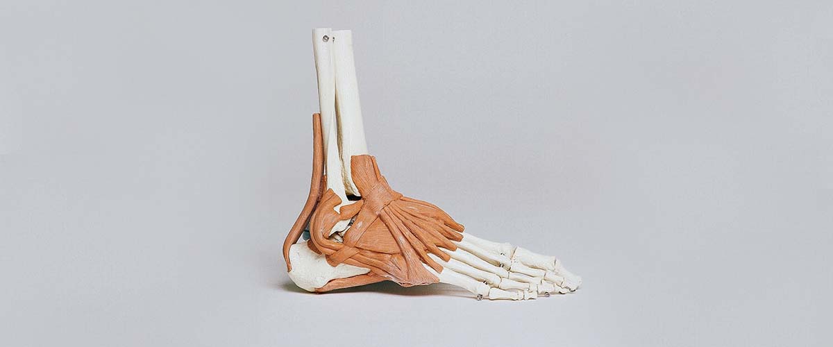
A Lisfranc injury is an injury to the midfoot, where the long metatarsal bones meet the small bones of the arch (the tarsometatarsal joints). It ranges from a sprain of the ligaments to fractures and dislocations.2 Most are not high-energy injuries: in one population study only 31% came from high-energy trauma,1 and many happen from a twist or a misstep, or when someone lands on a foot pointed downward. Because X-rays can look normal, subtle Lisfranc injuries are commonly overlooked.4
- Midfoot pain, swelling and difficulty bearing weight after a twist, fall or sports injury
- Bruising on the sole of the arch is a warning sign5
- Weight-bearing X-rays of both feet are key to diagnosis; CT or MRI for subtle injuries6,7
- Stable injuries usually heal in a boot without surgery12,13
- Unstable injuries need surgery: fixation or fusion, with a precise reduction being what matters most15,21
Why Lisfranc injuries matter
The Lisfranc joints form the keystone of the arch. When the ligaments that hold them together tear, the arch can shift and collapse, which can cause pain and arthritis. Missed or untreated Lisfranc injuries can cause long-term disability,2,4 which is why any midfoot pain after an injury deserves a careful check.3
Symptoms
- Pain and swelling on the top of the midfoot
- Difficulty or pain when standing on tiptoe or pushing off
- Bruising on the sole of the arch (the plantar ecchymosis sign)5
- Tenderness over the joints between the big-toe and second-toe metatarsals and the arch bones
- Pain with squeezing the forefoot or twisting the midfoot6
Diagnosis
Dr. Dold examines the foot for tenderness, swelling and pain with stress tests of the midfoot. Imaging is the key step:
- Weight-bearing X-rays of both feet: standing views show widening or shifting that non-weight-bearing views can miss, and comparing both feet helps detect subtle instability.6,7,8,9
- CT scan: shows alignment and small fractures hidden on X-rays.7 Weight-bearing CT, where available, detects subtle changes more accurately than conventional CT.11
- MRI: shows the ligaments themselves. In one study, MRI correctly classified 90% of injuries as stable or unstable.10
Stable injuries: treatment without surgery
When the joints are stable and lined up on weight-bearing imaging, treatment is a walking boot or cast, often with a period of limited weight-bearing, then a gradual return to activity. Results are good:
- Stable injuries treated without surgery had excellent outcomes at a median of 55 months.12
- Small avulsion fractures and simple fractures displaced less than 2 mm did well without surgery.13
- In a small randomized trial of non-displaced injuries, non-surgical care and surgery had similar 2-year scores.14
- Athletes with undisplaced or stable minimally displaced injuries all returned to sport after non-surgical treatment.30
Follow-up weight-bearing X-rays confirm the joint stays aligned while it heals.12
Unstable injuries: surgery
If the joints have shifted or are unstable, surgery is recommended to restore the arch. The single most important factor in the outcome is how accurately the joints are put back in place: anatomic reduction predicts better function and a lower risk of arthritis,15,16,17 and post-traumatic arthritis has been reported in about a third of patients after these injuries overall.17
The main options are:
- Open reduction and internal fixation (ORIF): realigning the joints and holding them with screws and/or a plate on top of the joints (bridge plating), which often comes out later. Dorsal bridge plates gave better scores than screws across the joints in one study,18 though implant type matters less than reduction quality.15
- Flexible fixation (suture-button devices): used for selected ligament-only injuries, with satisfactory early outcomes and return to activity, but lower-quality evidence so far.28,29
- Primary fusion: realigning and permanently fusing the inner two or three joints, which are normally stiff joints, so little useful motion is lost.
Fixation or fusion?
This is one of the more debated questions in foot surgery:
- In a randomized trial of ligament-only injuries, primary fusion had higher 2-year scores than fixation.20
- A more recent randomized trial of displaced injuries found similar 2-year results; both are viable options.21
- Meta-analyses suggest fusion may give better scores, but the evidence is limited, and some found no difference in revision or patient-reported outcomes.22,23,24,25
- Fixation leads to more planned hardware removal; when that is excluded, reoperation rates were about the same.19,26,27
- In athletes, return to sport did not differ significantly between the two,32 and a consensus of foot and ankle surgeons favored fixation for unstable ligament injuries in elite athletes.33
Dr. Dold recommends an approach based on the injury pattern (ligament vs fracture), how long ago it happened, your age and activity, and your goals.
Lisfranc injuries in athletes
Lisfranc injuries are well known in football and other cutting sports. Across studies, 93% to 94% of athletes returned to sport, and 74% to 88% to their pre-injury level.31 In NFL players, 81.8% to 92.9% returned at a median of about 11 months,34,35 and Lisfranc injuries found at the NFL Combine were linked to a worse draft position.36 These are serious injuries for any athlete, and a careful, staged return protects the result.
Missed or late-diagnosed injuries
If a Lisfranc injury is found weeks or months later, surgery can still help. Delayed fixation of missed low-energy injuries reduced pain, and patients returned to work and sport.37 For long-standing injuries with arthritis, fusion is often the more reliable option.4
Recovery
- Non-surgical: a boot or cast with protected weight-bearing, then a gradual return to shoes, often with an arch support.
- After surgery: a period of non-weight-bearing in a splint and boot, then progressive weight-bearing and physical therapy. Plates or screws across joints are sometimes removed later.19,26
- Return to sport: usually months rather than weeks; NFL players returned at a median of about 11 months.34,35
Dr. Dold reviews your expected timeline based on your injury and treatment.
Frequently asked questions
Is a Lisfranc injury just a sprain?
Some are sprains of the midfoot ligaments, but even a ‘sprain’ can make the joints unstable. Weight-bearing X-rays, and sometimes CT or MRI, show whether the joint is stable.
Can a Lisfranc injury heal without surgery?
Yes, if the joints are stable and aligned. Stable injuries treated in a boot had excellent outcomes in research studies. Unstable or displaced injuries generally need surgery.
How do I know if my Lisfranc injury was missed?
Ongoing midfoot pain, swelling or difficulty pushing off weeks after a foot injury, especially with bruising on the sole of the arch, should be evaluated with weight-bearing X-rays of both feet.
Will the screws or plate need to come out?
Implants that cross joints are often removed after healing. Fusion and flexible fixation usually avoid planned removal.
How long until I can play sports after a Lisfranc injury?
It depends on the injury and treatment. Most athletes return to sport, but usually months rather than weeks after an unstable injury.
Research cited on this page
- Stødle AH, Hvaal KH, Enger M, et al. Lisfranc injuries: Incidence, mechanisms of injury and predictors of instability. Foot Ankle Surg. 2020;26(5):535-540. PubMed
Lisfranc injuries occurred in 14 per 100,000 people per year, and only 31% were high-energy injuries. - Welck MJ, Zinchenko R, Rudge B. Lisfranc injuries. Injury. 2015;46(4):536-41. PubMed
Lisfranc injuries range from purely ligament injuries to fractures and are often difficult to diagnose; missed injuries can cause long-term disability. - McDermott A, Repanshek Z, Koyfman A, et al. High risk and low incidence diseases: Lisfranc injury. Am J Emerg Med. 2024;85:172-178. PubMed
Considering a Lisfranc injury helps emergency clinicians make a timely diagnosis and prevent complications. - Bowlby MA. Subtle Lisfranc Injuries. Clin Podiatr Med Surg. 2025;42(2):207-221. PubMed
Subtle Lisfranc injuries are commonly overlooked; midfoot pain after even minor trauma warrants bilateral weight-bearing X-rays. - Ross G, Cronin R, Hauzenblas J, et al. Plantar ecchymosis sign: a clinical aid to diagnosis of occult Lisfranc tarsometatarsal injuries. J Orthop Trauma. 1996;10(2):119-22. PubMed
Bruising on the sole of the midfoot (plantar ecchymosis sign) suggests a significant Lisfranc injury. - Semelsberger SD, Boggiano VJ, Webber K, et al. Diagnostic evaluation and nonoperative management of Lisfranc injuries in athletes. Knee Surg Sports Traumatol Arthrosc. 2026;34(9):3079-3088. PubMed
An international consensus recommended bilateral weight-bearing X-rays first, with CT or MRI for low-grade instability. - Sripanich Y, Weinberg MW, Krähenbühl N, et al. Imaging in Lisfranc injury: a systematic literature review. Skeletal Radiol. 2020;49(1):31-53. PubMed
Weight-bearing X-rays improve detection of subtle Lisfranc injuries; CT shows alignment and hidden fractures. - De Bruijn J, Hagemeijer NC, Rikken QGH, et al. Lisfranc injury: Refined diagnostic methodology using weightbearing and non-weightbearing radiographs. Injury. 2022;53(6):2318-2325. PubMed
Weight-bearing imaging revealed larger joint widening and was more reliable than non-weight-bearing imaging. - Rikken QGH, Hagemeijer NC, De Bruijn J, et al. Novel values in the radiographic diagnosis of ligamentous Lisfranc injuries. Injury. 2022;53(6):2326-2332. PubMed
Bilateral weight-bearing X-rays with a standardized measurement protocol can diagnose ligament-only Lisfranc instability. - Raikin SM, Elias I, Dheer S, et al. Prediction of midfoot instability in the subtle Lisfranc injury. Comparison of magnetic resonance imaging with intraoperative findings. J Bone Joint Surg Am. 2009;91(4):892-9. PubMed
MRI correctly classified 90% of Lisfranc joints as stable or unstable; plantar ligament disruption was the strongest predictor. - Talaski GM, Baumann AN, Walley KC, et al. Weightbearing Computed Tomography vs Conventional Tomography for Examination of Varying Degrees of Lisfranc Injures: A Systematic Review of the Literature. Foot Ankle Orthop. 2023;8(4):24730114231209767. PubMed
Weight-bearing CT measured the Lisfranc area more accurately and detected subtle changes better than conventional CT. - Stødle AH, Hvaal KH, Brøgger H, et al. Outcome after nonoperative treatment of stable Lisfranc injuries. A prospective cohort study. Foot Ankle Surg. 2022;28(2):245-250. PubMed
Stable Lisfranc injuries treated without surgery had excellent outcomes at a median of 55 months. - Ponkilainen VT, Partio N, Salonen EE, et al. Outcomes after nonoperatively treated non-displaced Lisfranc injury: a retrospective case series of 55 patients. Arch Orthop Trauma Surg. 2021;141(8):1311-1317. PubMed
Avulsion and simple fractures with less than 2 mm of displacement can be treated without surgery with good results. - Ponkilainen V, Mäenpää H, Laine HJ, et al. Operative versus non-operative treatment for non-displaced Lisfranc injuries: A two-center randomized clinical trial. Scand J Surg. 2025;114(2):153-161. PubMed
In a small randomized trial of non-displaced injuries, non-surgical care and surgery had similar 2-year scores. - Lau S, Guest C, Hall M, et al. Functional Outcomes Post Lisfranc Injury-Transarticular Screws, Dorsal Bridge Plating or Combination Treatment?. J Orthop Trauma. 2017;31(8):447-452. PubMed
The quality of anatomic reduction, not the type of implant, most strongly predicted function. - Stavrakakis I, Sperelakis I, Zampetakis K, et al. Outcomes and complications after open reduction and internal fixation of Lisfranc injuries: a retrospective cohort study. Eur J Orthop Surg Traumatol. 2025;35(1):271. PubMed
Anatomically reduced injuries had better scores and a lower risk of post-traumatic arthritis. - Tonsuthanluck S, Dahmen J, Kerkhoffs GMMJ, et al. Medial tarsometatarsal injuries lead to overall post-traumatic osteoarthritis in 1 in 3 patients: a systematic review and meta-analysis. Eur J Orthop Surg Traumatol. 2026;36(1). PubMed
Across 686 patients, post-traumatic arthritis developed in about 33%; non-anatomic reduction increased the risk. - Kirzner N, Zotov P, Goldbloom D, et al. Dorsal bridge plating or transarticular screws for Lisfranc fracture dislocations: a retrospective study comparing functional and radiological outcomes. Bone Joint J. 2018;100-B(4):468-474. PubMed
Dorsal bridge plating gave better functional scores than screws across the joints. - Ahmad J, Jones K. Randomized, Prospective Comparison of Bioabsorbable and Steel Screw Fixation of Lisfranc Injuries. J Orthop Trauma. 2016;30(12):676-681. PubMed
In a randomized trial, bioabsorbable and steel screws gave similar function and pain results. - Ly TV, Coetzee JC. Treatment of primarily ligamentous Lisfranc joint injuries: primary arthrodesis compared with open reduction and internal fixation. A prospective, randomized study. J Bone Joint Surg Am. 2006;88(3):514-20. PubMed
In a randomized trial of ligament-only injuries, primary fusion of the medial two or three rays had higher 2-year scores than screw fixation. - Ponkilainen V, Mäenpää H, Laine HJ, et al. Open Reduction Internal Fixation vs Primary Arthrodesis for Displaced Lisfranc Injuries: A Multicenter Randomized Controlled Trial. Foot Ankle Int. 2024;45(6):612-620. PubMed
In a randomized trial of displaced injuries, fixation and primary fusion had similar 2-year scores; both are viable. - Han PF, Zhang ZL, Chen CL, et al. Comparison of primary arthrodesis versus open reduction with internal fixation for Lisfranc injuries: Systematic review and meta-analysis. J Postgrad Med. 2019;65(2):93-100. PubMed
Meta-analysis: primary fusion had better scores and less hardware removal, with similar revision and complication rates. - Magill HHP, Hajibandeh S, Bennett J, et al. Open Reduction and Internal Fixation Versus Primary Arthrodesis for the Treatment of Acute Lisfranc Injuries: A Systematic Review and Meta-analysis. J Foot Ankle Surg. 2019;58(2):328-332. PubMed
Meta-analysis: primary fusion may give better pain and function, but the evidence was limited. - Smith N, Stone C, Furey A. Does Open Reduction and Internal Fixation versus Primary Arthrodesis Improve Patient Outcomes for Lisfranc Trauma? A Systematic Review and Meta-analysis. Clin Orthop Relat Res. 2016;474(6):1445-52. PubMed
Meta-analysis: fixation required more hardware removal; neither method was favored for revision or patient-reported outcomes. - Peters W, Panchbhavi V. Primary Arthrodesis Versus Open Reduction and Internal Fixation Outcomes for Lisfranc Injuries: An Analysis of Conflicting Meta-analyses Results. Foot Ankle Spec. 2022;15(2):171-178. PubMed
Primary fusion had lower hardware removal; most other outcomes did not differ. - Buda M, Kink S, Stavenuiter R, et al. Reoperation Rate Differences Between Open Reduction Internal Fixation and Primary Arthrodesis of Lisfranc Injuries. Foot Ankle Int. 2018;39(9):1089-1096. PubMed
Excluding planned hardware removal, reoperation rates were the same (about 30%) after fixation and fusion. - Henning JA, Jones CB, Sietsema DL, et al. Open reduction internal fixation versus primary arthrodesis for lisfranc injuries: a prospective randomized study. Foot Ankle Int. 2009;30(10):913-22. PubMed
Secondary surgery was 78.6% after fixation vs 16.7% after fusion, mostly planned hardware removal; function was similar. - O’Connor KP, Olfson ER, Riehl JT. Flexible fixation versus open reduction internal fixation and primary arthrodesis for ligamentous Lisfranc injuries: A systematic review and meta-analysis. Foot (Edinb). 2024;61:102145. PubMed
Flexible (suture-button) fixation had satisfactory outcomes, but the evidence was low quality. - Lachance AD, Giro ME, Edelstein A, et al. Suture button fixation yields high levels of patient reported outcomes, return to sport, and stable fixation in isolated Lisfranc injuries: A systematic review. J ISAKOS. 2023;8(6):474-483. PubMed
After suture-button fixation for isolated ligament injuries, all patients returned to sport or activity. - Robertson GAJ, Ang KK, Maffulli N, et al. Return to sport following Lisfranc injuries: A systematic review and meta-analysis. Foot Ankle Surg. 2019;25(5):654-664. PubMed
Non-displaced and stable minimally displaced injuries treated without surgery had a 100% return to sport. - Ter Laak Bolk CS, Dahmen J, Lambers KTA, et al. Adequate return to sports and sports activities after treatment of Lisfranc injury: a meta-analysis. J ISAKOS. 2021;6(4):212-219. PubMed
93% to 94% of athletes returned to sport after Lisfranc treatment, and 74% to 88% to their pre-injury level. - Feldman MW, Fucaloro S, Krivicich L, et al. Studies directly comparing Lisfranc injuries treated with primary arthrodesis or open reduction and internal fixation show no significant difference in return to sport and complications: A systematic review and meta-analysis. J Foot Ankle Surg. 2025;64(3):318-327. PubMed
Return to sport and complications did not differ significantly between fixation and primary fusion. - Balboni JM, Levine AR, Boggiano VJ, et al. Operative treatment of Lisfranc injuries in elite athletes: 2024 International Foot and Ankle Sports Consensus and systematic review. Knee Surg Sports Traumatol Arthrosc. 2026;34(9):3089-3105. PubMed
An international consensus recommended fixation rather than fusion for unstable ligament injuries in elite athletes. - McHale KJ, Rozell JC, Milby AH, et al. Outcomes of Lisfranc Injuries in the National Football League. Am J Sports Med. 2016;44(7):1810-7. PubMed
92.9% of NFL players returned to play at a median of 11.1 months after a Lisfranc injury. - Abed V, Fine R, Fine R, et al. Return to Play, Performance, and Economic Analysis of National Football League Players After Lisfranc Injury. Orthop J Sports Med. 2023;11(4):23259671231159935. PubMed
81.8% of NFL players returned at a median of 11 months, with a drop in performance at 1 year. - McHale KJ, Vopat BG, Beaulieu-Jones BR, et al. Epidemiology and Outcomes of Lisfranc Injuries Identified at the National Football League Scouting Combine. Am J Sports Med. 2017;45(8):1901-1908. PubMed
Lisfranc injuries found at the NFL Combine were linked to worse draft position. - Cassinelli SJ, Moss LK, Lee DC, et al. Delayed Open Reduction Internal Fixation of Missed, Low-Energy Lisfranc Injuries. Foot Ankle Int. 2016;37(10):1084-1090. PubMed
Delayed fixation of missed low-energy Lisfranc injuries reduced pain, and patients returned to work and sport.
Medically reviewed by Andrew P. Dold, MD, FAOA, FAAOS, FACS, FRCSC. Last reviewed October 2026. This page is general information, not medical advice. Your own diagnosis and treatment plan depend on your exam and imaging.