








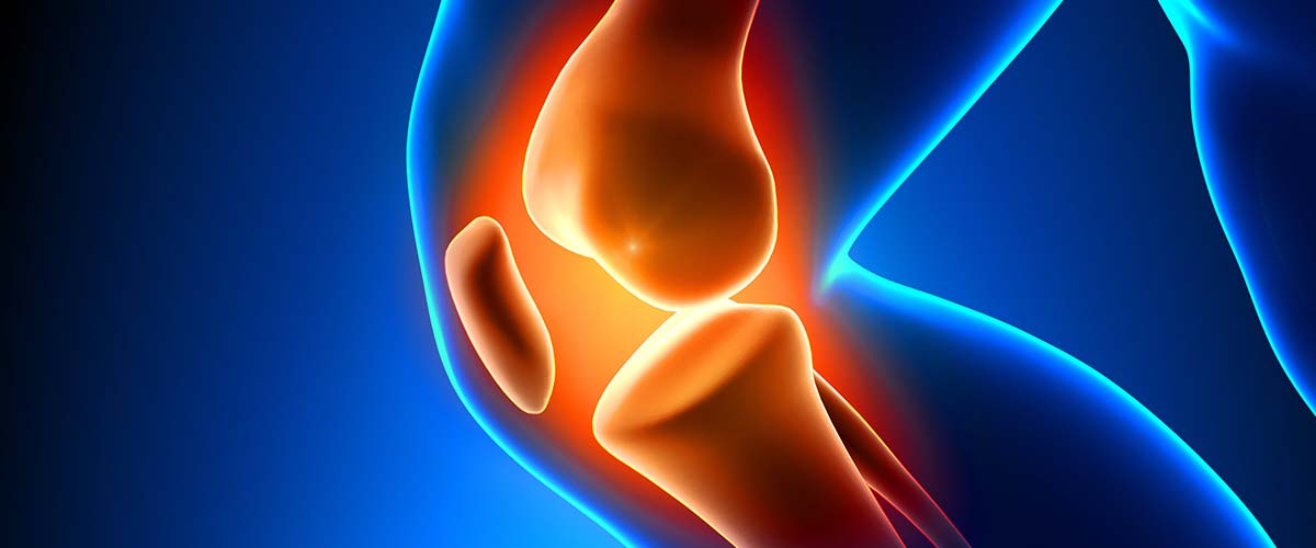
Osteonecrosis of the knee means an area of bone under the joint surface has lost its normal blood supply or structure. There are two main types. The more common one, spontaneous osteonecrosis of the knee (SONK), usually causes sudden pain in adults over 50 and is now understood to be a subchondral insufficiency fracture: a small fracture under the cartilage in weakened bone.1,2 Secondary osteonecrosis (avascular necrosis, AVN) tends to affect younger people, often after corticosteroid use, and frequently involves several areas.4,17,18
- SONK: sudden inner-knee pain, often at night, in older adults; usually linked to a meniscus root tear5,7
- Early treatment: protected weight-bearing and offloading; many small lesions settle2,14
- Large lesions, varus alignment and arthritis predict progression13,14,15
- Repairing a torn meniscus root can stop early insufficiency fractures from progressing11
- Collapse or arthritis: partial or total knee replacement28,31
SONK and insufficiency fractures
SONK most often affects the inner (medial) femoral condyle and is more common in women.4,5 In one large series of 223 patients, 71% were women, and 74% had a meniscal root or radial tear.5 When the meniscus root tears, the meniscus can no longer spread load, so the bone underneath is overloaded and can develop a small fracture:
- About 90% of patients with medial SONK had a tear of the same meniscus, and about 62% a posterior root tear.7
- Most studies implicate meniscal tears or meniscectomy in SONK,8,10 and osteonecrosis was more common in knees with root tears.9
Rarely, osteonecrosis is found after knee arthroscopy or ligament surgery.20,21
Secondary osteonecrosis (AVN)
Secondary osteonecrosis typically affects people under 50 to 55, often in both knees and other joints.4,17 Risk factors include corticosteroid use (75% of patients in one series), lupus and other immune conditions, blood disorders and heavy alcohol use.17,18 It often progresses, so earlier treatment may be considered.19
Symptoms
- Sudden, sharp pain on the inside of the knee, often without a clear injury
- Pain at night or at rest
- Tenderness over the inner thighbone
- Swelling and pain with weight-bearing
- With secondary AVN: more gradual pain, sometimes in both knees
Diagnosis
X-rays are often normal early on. MRI is the key test, showing bone marrow edema, the fracture line and the size of the lesion, as well as the meniscus root.3 Dr. Dold also measures leg alignment, because lesion size and varus (bow-legged) alignment strongly influence prognosis:
- A lesion deeper than 20 mm on MRI and a more varus knee predicted a poorer outcome.13
- Six of 7 patients with large lesions needed major surgery, while none of 10 with small lesions did.14
- Moderate-to-severe varus alignment was linked to a 13-fold higher risk of knee replacement.15
- Meniscus extrusion and existing arthritis also predict progression; 27% to 34% of patients progressed to surgery within a few years.5,6
Non-surgical treatment
Many small, early lesions improve with time:2,23
- Protected weight-bearing on crutches, often for about 3 to 6 weeks, sometimes with an unloader brace2,23
- Pain control and anti-inflammatory medicines when safe
- Bisphosphonates have been used; in a small series, 12% needed surgery vs 32% in an earlier untreated group, but randomized trials are lacking.16
- Checking bone health, since insufficiency fractures occur in weakened bone
Joint-preserving surgery
When pain persists or the lesion is at risk of progressing, and before the joint surface collapses, options include:22
- Meniscus root repair: repairing a torn medial meniscus root prevented progression of low-grade insufficiency fractures and improved outcomes.11 Without repair, 31% of patients with root tears had knee replacement at about 2.5 years.12
- Core decompression or subchondroplasty for bone marrow edema that does not settle.23,24
- High tibial osteotomy: shifting weight away from the damaged inner side of a varus knee, sometimes with drilling of the lesion.25
- Osteochondral allograft: replacing the damaged bone and cartilage in younger patients. Results were durable for SONK26 and for steroid-associated osteonecrosis, with 82% graft survival at 10 years.27
Joint-preserving surgery kept 87% of knees at a mean of 40 months in one series.24
Knee replacement
When the joint surface has collapsed or arthritis has developed, joint replacement is usually needed.22
- Partial (unicompartmental) knee replacement for SONK limited to one compartment: revision rates were about 5.5% at 6 years,28 survivorship was 92% at 15 years,29 and long-term results matched those for osteoarthritis.30 Total and partial replacement gave equivalent results in one comparison.31
- Total knee replacement when more of the knee is involved or for secondary osteonecrosis, with 92% survivorship free from revision in one series.32 Total knee replacement is the most common operation for knee osteonecrosis overall.33
Recovery
- Non-surgical: several weeks of protected weight-bearing, then gradual return to activity guided by symptoms and follow-up imaging.
- After root repair or osteotomy: a period of protected weight-bearing in a brace, then physical therapy over several months.
- After knee replacement: walking with support right away and progressive therapy.
Dr. Dold reviews your expected timeline based on your treatment.
Frequently asked questions
What is the difference between SONK and avascular necrosis?
SONK usually affects one area in an older adult and is now understood to be a small insufficiency fracture under the cartilage, often linked to a meniscus root tear. Secondary AVN tends to affect younger people, often after steroid use, and may involve several joints.
Will SONK heal on its own?
Many small lesions improve with protected weight-bearing over weeks to months. Larger lesions and those with collapse or arthritis are more likely to need surgery.
Why does a meniscus root tear matter?
The root anchors the meniscus. When it tears, the meniscus can’t spread load, and the bone underneath can be overloaded and fracture. Repairing the root early can help protect the bone.
Do I need a knee replacement?
Not always. Early, small lesions are often treated without surgery, and some patients benefit from root repair, osteotomy or grafting. If the joint has collapsed, a partial or total knee replacement gives reliable relief.
Should I worry about my other knee?
With secondary osteonecrosis, both knees and other joints are often involved, so Dr. Dold may recommend checking them. SONK usually affects one knee.
Research cited on this page
- Yamamoto T, Bullough PG. Spontaneous osteonecrosis of the knee: the result of subchondral insufficiency fracture. J Bone Joint Surg Am. 2000;82(6):858-66. PubMed
Tissue studies showed that what was called spontaneous osteonecrosis of the knee is the result of a subchondral insufficiency fracture. - Sibilska A, Góralczyk A, Hermanowicz K, et al. Spontaneous osteonecrosis of the knee: what do we know so far? A literature review. Int Orthop. 2020;44(6):1063-1069. PubMed
Early SONK appears to result from a subchondral fracture; non-surgical care includes protected weight-bearing, anti-inflammatories and bisphosphonates. - Ochi J, Nozaki T, Nimura A, et al. Subchondral insufficiency fracture of the knee: review of current concepts and radiological differential diagnoses. Jpn J Radiol. 2022;40(5):443-457. PubMed
Meniscal problems, including posterior root tears, are found in many patients with subchondral insufficiency fracture and are thought to contribute. - Lerebours F, ElAttrache NS, Mandelbaum B. Diseases of Subchondral Bone 2. Sports Med Arthrosc Rev. 2016;24(2):50-5. PubMed
SONK typically affects older women at the inner femoral condyle; secondary osteonecrosis affects people under 55 with multiple areas. - Pareek A, Parkes CW, Bernard C, et al. Spontaneous Osteonecrosis/Subchondral Insufficiency Fractures of the Knee: High Rates of Conversion to Surgical Treatment and Arthroplasty. J Bone Joint Surg Am. 2020;102(9):821-829. PubMed
In 223 patients (71% female), 74% had meniscal root or radial tears, and 34% progressed to surgery at about 2.7 years. - Pareek A, Parkes CW, Bernard CD, et al. The SIFK score: a validated predictive model for arthroplasty progression after subchondral insufficiency fractures of the knee. Knee Surg Sports Traumatol Arthrosc. 2020;28(10):3149-3155. PubMed
27% needed knee replacement at about 4 years; meniscus extrusion and existing arthritis predicted progression. - Feng Y, Yang J, Zhang M, et al. Association of Spontaneous Osteonecrosis of the Knee with Ipsilateral Meniscus and Posterior Root Tear: Systematic Review and Meta-analysis. Indian J Orthop. 2024;58(9):1188-1195. PubMed
In medial SONK, about 90% had a tear of the same-side meniscus and about 62% a posterior root tear. - Hussain ZB, Chahla J, Mandelbaum BR, et al. The Role of Meniscal Tears in Spontaneous Osteonecrosis of the Knee: A Systematic Review of Suspected Etiology and a Call to Revisit Nomenclature. Am J Sports Med. 2019;47(2):501-507. PubMed
21 of 26 studies implicated meniscal tears or meniscectomy in the development of SONK. - Sung JH, Ha JK, Lee DW, et al. Meniscal extrusion and spontaneous osteonecrosis with root tear of medial meniscus: comparison with horizontal tear. Arthroscopy. 2013;29(4):726-32. PubMed
Osteonecrosis occurred in 33.3% of knees with a medial meniscus root tear vs 14.8% with other tears. - Muscolo DL, Costa-Paz M, Ayerza M, et al. Medial meniscal tears and spontaneous osteonecrosis of the knee. Arthroscopy. 2006;22(4):457-60. PubMed
MRI documented degenerative medial meniscal tears followed by spontaneous osteonecrosis without any surgery. - Okazaki Y, Furumatsu T, Hiranaka T, et al. Medial meniscus posterior root repair prevents the progression of subchondral insufficiency fracture of the knee. J Orthop Sci. 2021;26(6):1051-1055. PubMed
Repairing the medial meniscus root prevented progression of low-grade insufficiency fractures and improved outcomes. - Krych AJ, Reardon PJ, Johnson NR, et al. Non-operative management of medial meniscus posterior horn root tears is associated with worsening arthritis and poor clinical outcome at 5-year follow-up. Knee Surg Sports Traumatol Arthrosc. 2017;25(2):383-389. PubMed
With non-surgical care of medial meniscus root tears, 31% had knee replacement at a mean of 30 months. - Akamatsu Y, Kobayashi H, Kusayama Y, et al. Predictive factors for the progression of spontaneous osteonecrosis of the knee. Knee Surg Sports Traumatol Arthrosc. 2017;25(2):477-484. PubMed
A more varus knee and a lesion deeper than 20 mm on MRI predicted a poorer prognosis. - Juréus J, Lindstrand A, Geijer M, et al. The natural course of spontaneous osteonecrosis of the knee (SPONK): a 1- to 27-year follow-up of 40 patients. Acta Orthop. 2013;84(4):410-4. PubMed
6 of 7 patients with large lesions needed major surgery, while none of 10 with small lesions did. - Park JY, Cho BW, Kim TH, et al. Moderate-to-Severe Varus Deformity Is Associated With Conversion to Arthroplasty in Patients Who Have Subchondral Insufficiency Fracture of the Knee. J Arthroplasty. 2026;41(9):2717-2722.e2. PubMed
Moderate-to-severe varus alignment was linked to a 13-fold higher risk of needing knee replacement. - Jureus J, Lindstrand A, Geijer M, et al. Treatment of spontaneous osteonecrosis of the knee (SPONK) by a bisphosphonate. Acta Orthop. 2012;83(5):511-4. PubMed
In a small series treated with a bisphosphonate, 12% needed surgery vs 32% in an earlier untreated series. - Mont MA, Baumgarten KM, Rifai A, et al. Atraumatic osteonecrosis of the knee. J Bone Joint Surg Am. 2000;82(9):1279-90. PubMed
In atraumatic (secondary) osteonecrosis, the mean age was 36, 74% had other joints involved and most had immune-related disease. - Boontanapibul K, Steere JT, Amanatullah DF, et al. Initial Presentation and Progression of Secondary Osteonecrosis of the Knee. J Arthroplasty. 2020;35(10):2798-2806. PubMed
In secondary knee osteonecrosis, 75% had used corticosteroids and 58% had both knees affected. - Mont MA, Marker DR, Zywiel MG, et al. Osteonecrosis of the knee and related conditions. J Am Acad Orthop Surg. 2011;19(8):482-94. PubMed
Secondary osteonecrosis often progresses to end-stage disease; core decompression, bone grafting, osteotomy and arthroplasty are used. - Türker M, Çetik Ö, Çırpar M, et al. Postarthroscopy osteonecrosis of the knee. Knee Surg Sports Traumatol Arthrosc. 2015;23(1):246-50. PubMed
Osteonecrosis after arthroscopy was uncommon; adding chondroplasty to meniscectomy did not increase it. - Lansdown DA, Shaw J, Allen CR, et al. Osteonecrosis of the Knee After Anterior Cruciate Ligament Reconstruction: A Report of 5 Cases. Orthop J Sports Med. 2015;3(3):2325967115576120. PubMed
Osteonecrosis after ACL reconstruction is rare and was diagnosed at a mean of 11.6 months. - Karim AR, Cherian JJ, Jauregui JJ, et al. Osteonecrosis of the knee: review. Ann Transl Med. 2015;3(1):6. PubMed
Joint-preserving procedures work in lesions before collapse; after severe collapse, joint replacement is usually needed. - Villari E, Digennaro V, Panciera A, et al. Bone marrow edema of the knee: a narrative review. Arch Orthop Trauma Surg. 2024;144(5):2305-2316. PubMed
Bone marrow edema is first treated with 3 to 6 weeks off weight-bearing; subchondroplasty and core decompression are options for non-responders. - Duany NG, Zywiel MG, McGrath MS, et al. Joint-preserving surgical treatment of spontaneous osteonecrosis of the knee. Arch Orthop Trauma Surg. 2010;130(1):11-6. PubMed
Joint-preserving surgery (core decompression or osteochondral grafting) kept 87% of knees at a mean of 40 months. - Takeuchi R, Aratake M, Bito H, et al. Clinical results and radiographical evaluation of opening wedge high tibial osteotomy for spontaneous osteonecrosis of the knee. Knee Surg Sports Traumatol Arthrosc. 2009;17(4):361-8. PubMed
Drilling with an opening-wedge high tibial osteotomy relieved pain in SONK. - Tírico LEP, Early SA, McCauley JC, et al. Fresh Osteochondral Allograft Transplantation for Spontaneous Osteonecrosis of the Knee: A Case Series. Orthop J Sports Med. 2017;5(10):2325967117730540. PubMed
Fresh osteochondral allografts for stage 2 and 3 SONK gave durable results with no graft failures. - Early S, Tírico LEP, Pulido PA, et al. Long-Term Retrospective Follow-Up of Fresh Osteochondral Allograft Transplantation for Steroid-Associated Osteonecrosis of the Femoral Condyles. Cartilage. 2021;12(1):24-30. PubMed
Osteochondral allografts for steroid-associated osteonecrosis had 82% survival at 10 years; 85% avoided arthroplasty. - Jauregui JJ, Blum CL, Sardesai N, et al. Unicompartmental knee arthroplasty for spontaneous osteonecrosis of the knee: A meta-analysis. J Orthop Surg (Hong Kong). 2018;26(2):2309499018770925. PubMed
Partial (unicompartmental) knee replacement for SONK had a revision rate of about 5.5% at a mean of 6 years. - Ollivier M, Jacquet C, Lucet A, et al. Long-Term Results of Medial Unicompartmental Knee Arthroplasty for Knee Avascular Necrosis. J Arthroplasty. 2019;34(3):465-468. PubMed
Partial knee replacement for osteonecrosis had 92% survivorship at 15 years. - Ly L, Batailler C, Shatrov J, et al. Satisfactory Outcomes of All-Poly Fixed Bearing Unicompartmental Knee Arthroplasty for Avascular Osteonecrosis Versus Osteoarthritis: A Comparative Study With 10 to 22 Years of Follow-up. J Arthroplasty. 2022;37(9):1743-1750. PubMed
At 10 to 21 years, partial knee replacement for SONK had results similar to osteoarthritis and 85.1% survival free from revision. - Flury A, Weigelt L, Camenzind RS, et al. Total and unicondylar knee arthroplasty are equivalent treatment options in end-stage spontaneous osteonecrosis of the knee, and the size of the lesion has no influence on the results. Knee Surg Sports Traumatol Arthrosc. 2021;29(10):3254-3261. PubMed
Total and partial knee replacement for SONK had equivalent results, with slightly better function after partial replacement. - Boontanapibul K, Amanatullah DF, Huddleston JI, et al. Outcomes of Cemented Total Knee Arthroplasty for Secondary Osteonecrosis of the Knee. J Arthroplasty. 2021;36(2):550-559. PubMed
Cemented total knee replacement for secondary osteonecrosis had 92% survivorship free from revision. - Ng MK, Gordon AM, Lam A, et al. Surgical Trends for Managing Knee Osteonecrosis: A 2010 to 2020 United States Nationwide Study. J Arthroplasty. 2023;38(6S):S125-S130. PubMed
Total knee replacement accounted for 85.4% of surgery for knee osteonecrosis; joint-preserving surgery was more common under age 50.
Medically reviewed by Andrew P. Dold, MD, FAOA, FAAOS, FACS, FRCSC. Last reviewed October 2026. This page is general information, not medical advice. Your own diagnosis and treatment plan depend on your exam and imaging.