








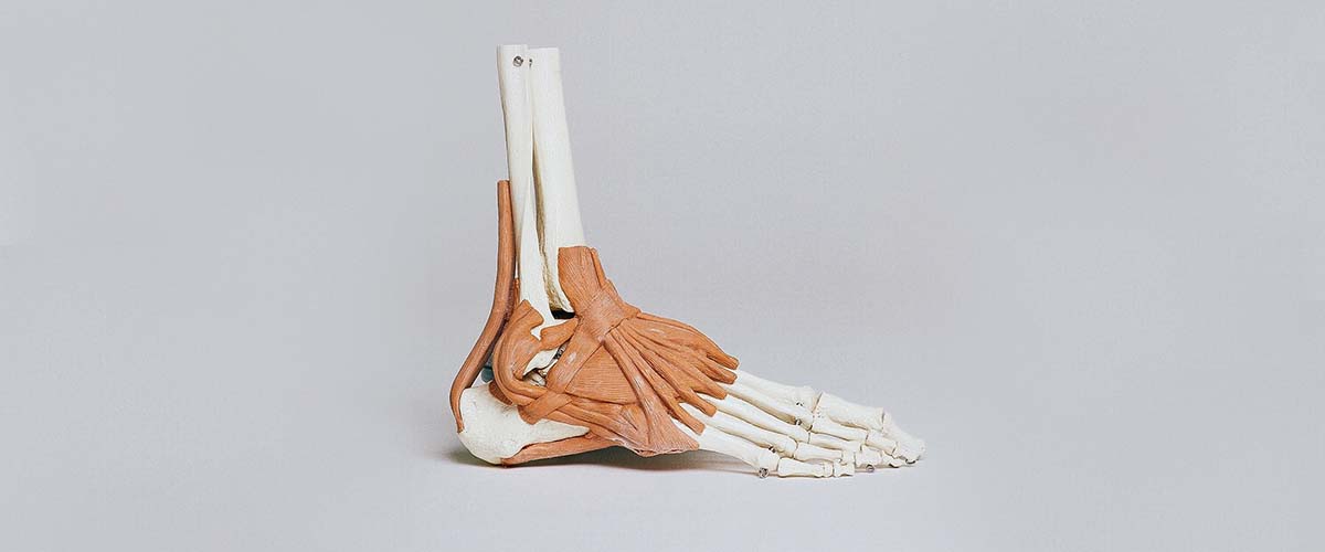
An osteochondral lesion of the talus (OLT), sometimes called talar OCD or a talar dome lesion, is damage to the cartilage and the bone just beneath it on the top of the talus, the bone that forms the lower half of the ankle joint.1 Many follow an ankle sprain or fracture: in athletes with an acute ankle injury, MRI showed a cartilage or osteochondral lesion in 14%.3 These injuries are a common reason an ankle “sprain” keeps hurting months later.
- Deep, aching ankle pain, swelling or catching that lingers after a sprain
- Often missed on X-ray; MRI or CT shows the lesion
- Non-surgical care helps some patients, especially children4,6,7
- Small lesions: arthroscopic bone marrow stimulation (microfracture)9,10,11
- Larger or failed lesions: cartilage and bone grafting20,22
Who gets OLTs?
OLTs mostly affect active adults, often men in their thirties.2 There are two common patterns:
- Lateral (outside) lesions: usually smaller and shallower, often after an ankle sprain, and frequently with ligament damage.2
- Medial (inside) lesions: more common, wider and deeper, and often without a clear injury.2
In children and teens, the condition is often called juvenile osteochondritis dissecans, and stable lesions in this age group are more likely to heal without surgery.4
Symptoms
- Deep ankle pain with walking, running or jumping, often weeks or months after a sprain
- Swelling after activity
- Catching, locking or a feeling of instability
- Stiffness
Diagnosis
X-rays can look normal. Dr. Dold orders an MRI to see the cartilage, bone bruising and any loose fragment, and a CT scan when the bone and the size and depth of the lesion need to be measured for surgical planning. Lesion size, depth, location, whether the fragment is loose, and whether cysts are present all guide treatment. The ankle ligaments are checked too, since instability often goes along with lateral lesions.2
Non-surgical treatment
For stable lesions and new injuries, treatment starts with a period of rest, often in a boot with protected weight-bearing, followed by physical therapy and a gradual return to activity. Results vary:
- Across 868 patients treated without surgery, about 45% had a successful outcome, and arthritis progressed in 9%.6
- Some patients improve substantially in pain and function while the lesion itself stays the same size.5
- Children and teens do better with conservative care than adults.4,7
Injections for OLT have little evidence: few studies exist, and hyaluronic acid and PRP did not differ in the available comparison.8
Arthroscopic bone marrow stimulation (microfracture)
For small lesions that keep hurting despite non-surgical care, the most common first operation is arthroscopic debridement and bone marrow stimulation (microfracture or drilling): damaged cartilage is removed and small holes are made in the bone so healing cells can form repair tissue.9
Size matters:
- In one study, lesions of 150 mm² or more had an 80% failure rate after marrow stimulation, vs 10.5% for smaller lesions.11
- A systematic review linked outcome to lesion size in studies averaging about 107 mm²,10 and newer data suggest marrow stimulation is reserved for lesions smaller than about 107 mm².12
- At 10 years, 18% needed reoperation; obesity, age over 40 and existing arthritis worsened results.13
In athletes, return to activity after microfracture or bone grafting averaged about 17 weeks.14
Biologic augmentation
Several add-ons aim to improve the repair tissue after microfracture:
- PRP: in a randomized trial, adding PRP to microfracture gave better function and pain scores than microfracture alone.15
- Bone marrow aspirate concentrate (BMAC): revision rates were 28.8% after microfracture alone vs 12.2% with BMAC in one study.16
- Juvenile cartilage grafts: results have been mixed in small studies.17,18
These studies are generally small, so Dr. Dold discusses the strength of the evidence with each patient.
Larger, deeper or failed lesions
- Osteochondral autograft transfer (OATS or mosaicplasty): plugs of healthy cartilage and bone, usually from a non-weight-bearing area of the knee, are moved into the defect. After autograft transfer, 86.3% of athletes returned to play, at a mean of 5.8 months,20 with favorable long-term results.21 Mosaicplasty relieved pain more than microfracture in one comparison.19
- Fresh osteochondral allograft: donor cartilage and bone for large lesions, with graft survival of 86.6% at intermediate follow-up,22 though 25% needed another operation in one review.23
- Cartilage cell and membrane techniques (ACI, MACI, AMIC): used for larger defects, with improved outcomes but low-quality evidence so far.24,25
- Retrograde drilling: for lesions with cysts under intact cartilage, drilling from below the joint avoids damaging the surface.26
- Fragment fixation: a loose but healthy fragment can sometimes be fixed back in place with pins or screws.31,32
Medial lesions at the back of the talus sometimes require cutting and later fixing the inner ankle bone (medial malleolar osteotomy) for access; in one series it healed in 66 of 67 patients.29 Long-term, survival without revision has been high for most cartilage techniques.27
Recovery and return to sport
- After microfracture: a period of protected or non-weight-bearing, early ankle motion, then strengthening and a gradual return to impact.
- After grafting or osteotomy: usually about 5 to 6 weeks of non-weight-bearing; one study found no difference between 5 and 6 weeks.30
- Return to sport: in one prospective study, 70.6% of athletes returned, at an average of 4.3 months.28
A second arthroscopy for a lesion that fails often gives only partial relief,33 which is why matching the first operation to the lesion matters.
Frequently asked questions
Will an OLT heal on its own?
Some do, especially in children and teens with stable lesions. In adults, about half of patients do well without surgery; the rest may need an arthroscopic procedure.
Why did my ankle sprain never get better?
Persistent deep pain, swelling or catching months after a sprain can come from a cartilage injury on the talus, an unstable ligament or both. An MRI usually shows the problem.
What is microfracture?
An arthroscopic procedure that clears damaged cartilage and makes small holes in the bone so the body can fill the defect with repair tissue. It works well for smaller lesions.
What if my lesion is large?
Larger or deeper lesions, and those that fail microfracture, may be treated with cartilage and bone grafts from your knee or a donor, or with cartilage cell techniques.
How long until I can run again?
It depends on the procedure. In studies, athletes returned to sport about 4 to 6 months after surgery on average.
Research cited on this page
- Murawski CD, Jamal MS, Hurley ET, et al. Terminology for osteochondral lesions of the ankle: proceedings of the International Consensus Meeting on Cartilage Repair of the Ankle. J ISAKOS. 2022;7(2):62-66. PubMed
An international consensus defined standard terms for osteochondral lesions of the ankle, including OLT and bone marrow stimulation. - Barbier O, Amouyel T, de l’Escalopier N, et al. Osteochondral lesion of the talus: What are we talking about?. Orthop Traumatol Surg Res. 2021;107(8S):103068. PubMed
OLTs mostly affect men in their thirties; lateral lesions tend to be smaller and follow trauma, medial lesions are larger and deeper. - Baltes TPA, Dalansi F, Al-Naimi MR, et al. The Prevalence, Size, and Anatomic Location of Cartilage and Osteochondral Lesions in Athletes With an Acute Ligamentous Ankle Injury. Am J Sports Med. 2025;53(9):2173-2180. PubMed
On MRI after acute ankle injuries in athletes, 14% had cartilage or osteochondral lesions. - Bruns J, Habermann C, Werner M. Osteochondral Lesions of the Talus: A Review on Talus Osteochondral Injuries, Including Osteochondritis Dissecans. Cartilage. 2021;13(1_suppl):1380S-1401S. PubMed
Stable, non-displaced lesions in children can often be treated without surgery; this is less successful in adults. - Seo SG, Kim JS, Seo DK, et al. Osteochondral lesions of the talus. Acta Orthop. 2018;89(4):462-467. PubMed
Many patients treated without surgery improved in pain and function, and lesion size usually did not change. - Buck TMF, Lauf K, Dahmen J, et al. Non-operative management for osteochondral lesions of the talus: a systematic review of treatment modalities, clinical- and radiological outcomes. Knee Surg Sports Traumatol Arthrosc. 2023;31(8):3517-3527. PubMed
Across 868 patients treated without surgery, the pooled success rate was 45%; arthritis progressed in 9%. - Dahmen J, Steman JAH, Buck TMF, et al. Treatment of Osteochondral Lesions of the Talus in the Skeletally Immature Population: A Systematic Review. J Pediatr Orthop. 2022;42(8):e852-e860. PubMed
In children and teens, success was 44% with conservative care and 77% after bone marrow stimulation. - Boffa A, Previtali D, Di Laura Frattura G, et al. Evidence on ankle injections for osteochondral lesions and osteoarthritis: a systematic review and meta-analysis. Int Orthop. 2021;45(2):509-523. PubMed
For OLT, injection studies were few; hyaluronic acid and PRP did not differ, and evidence was very low. - Zengerink M, Struijs PA, Tol JL, et al. Treatment of osteochondral lesions of the talus: a systematic review. Knee Surg Sports Traumatol Arthrosc. 2010;18(2):238-46. PubMed
Bone marrow stimulation was identified as an effective first-line surgical treatment for primary talar lesions. - Ramponi L, Yasui Y, Murawski CD, et al. Lesion Size Is a Predictor of Clinical Outcomes After Bone Marrow Stimulation for Osteochondral Lesions of the Talus: A Systematic Review. Am J Sports Med. 2017;45(7):1698-1705. PubMed
Lesion size predicted outcome after bone marrow stimulation in studies with a mean lesion area of about 107 mm². - Choi WJ, Park KK, Kim BS, et al. Osteochondral lesion of the talus: is there a critical defect size for poor outcome?. Am J Sports Med. 2009;37(10):1974-80. PubMed
Lesions of 150 mm² or more had an 80% failure rate after marrow stimulation, vs 10.5% for smaller lesions. - Kimball JS, Ferkel RD, Ferkel EI. Regeneration: Bone-Marrow Stimulation of the Talus-Limits and Goals. Foot Ankle Clin. 2024;29(2):281-290. PubMed
Newer data suggest bone marrow stimulation is reserved for lesions smaller than about 107 mm². - Hankemeier S, Müller EJ, Kaminski A, et al. [10-year results of bone marrow stimulating therapy in the treatment of osteochondritis dissecans of the talus]. Unfallchirurg. 2003;106(6):461-6. PubMed
At 10 years after marrow stimulation, 18% needed reoperation; obesity, age over 40 and existing arthritis worsened results. - Saxena A, Eakin C. Articular talar injuries in athletes: results of microfracture and autogenous bone graft. Am J Sports Med. 2007;35(10):1680-7. PubMed
In athletes, microfracture and bone grafting both improved scores; return to activity averaged about 17 weeks. - Guney A, Akar M, Karaman I, et al. Clinical outcomes of platelet rich plasma (PRP) as an adjunct to microfracture surgery in osteochondral lesions of the talus. Knee Surg Sports Traumatol Arthrosc. 2015;23(8):2384-2389. PubMed
In a randomized trial, adding PRP to microfracture gave better function and pain scores than microfracture alone. - Murphy EP, McGoldrick NP, Curtin M, et al. A prospective evaluation of bone marrow aspirate concentrate and microfracture in the treatment of osteochondral lesions of the talus. Foot Ankle Surg. 2019;25(4):441-448. PubMed
Revision rates were 28.8% after microfracture alone vs 12.2% when bone marrow aspirate concentrate was added. - Karnovsky SC, DeSandis B, Haleem AM, et al. Comparison of Juvenile Allogenous Articular Cartilage and Bone Marrow Aspirate Concentrate Versus Microfracture With and Without Bone Marrow Aspirate Concentrate in Arthroscopic Treatment of Talar Osteochondral Lesions. Foot Ankle Int. 2018;39(4):393-405. PubMed
Juvenile cartilage with bone marrow concentrate and microfracture gave similar patient-reported outcomes. - Lanham NS, Carroll JJ, Cooper MT, et al. A Comparison of Outcomes of Particulated Juvenile Articular Cartilage and Bone Marrow Aspirate Concentrate for Articular Cartilage Lesions of the Talus. Foot Ankle Spec. 2017;10(4):315-321. PubMed
In a small comparison, particulated juvenile cartilage had higher scores than a bone-marrow-based technique. - Guney A, Yurdakul E, Karaman I, et al. Medium-term outcomes of mosaicplasty versus arthroscopic microfracture with or without platelet-rich plasma in the treatment of osteochondral lesions of the talus. Knee Surg Sports Traumatol Arthrosc. 2016;24(4):1293-8. PubMed
Mosaicplasty and microfracture gave good mid-term results; pain improved more after mosaicplasty. - Seow D, Shimozono Y, Gianakos AL, et al. Autologous osteochondral transplantation for osteochondral lesions of the talus: high rate of return to play in the athletic population. Knee Surg Sports Traumatol Arthrosc. 2021;29(5):1554-1561. PubMed
After osteochondral autograft transfer, 86.3% of athletes returned to play at a mean of 5.8 months. - Winkler PW, Geyer S, Walzl D, et al. Favorable long-term clinical and radiologic outcomes with high survivorship after autologous osteochondral transplantation of the talus. Knee Surg Sports Traumatol Arthrosc. 2023;31(6):2166-2173. PubMed
Osteochondral autograft transfer showed favorable long-term clinical and MRI results without donor-site problems. - Pereira GF, Steele JR, Fletcher AN, et al. Fresh Osteochondral Allograft Transplantation for Osteochondral Lesions of the Talus: A Systematic Review. J Foot Ankle Surg. 2021;60(3):585-591. PubMed
Fresh osteochondral allograft had an aggregate graft survival of 86.6% at intermediate follow-up. - VanTienderen RJ, Dunn JC, Kusnezov N, et al. Osteochondral Allograft Transfer for Treatment of Osteochondral Lesions of the Talus: A Systematic Review. Arthroscopy. 2017;33(1):217-222. PubMed
After talar allograft, scores improved, but 25% of patients needed at least one more operation. - Erickson B, Fillingham Y, Hellman M, et al. Surgical management of large talar osteochondral defects using autologous chondrocyte implantation. Foot Ankle Surg. 2018;24(2):131-136. PubMed
Chondrocyte implantation for large talar defects improved outcomes, but the evidence was low quality. - Migliorini F, Maffulli N, Baroncini A, et al. Matrix-induced autologous chondrocyte implantation versus autologous matrix-induced chondrogenesis for chondral defects of the talus: a systematic review. Br Med Bull. 2021;138(1):144-154. PubMed
Two cell-and-membrane cartilage techniques (AMIC and MACI) gave similar clinical results. - Artioli E, Mazzotti A, Gerardi S, et al. Retrograde drilling for ankle joint osteochondral lesions: a systematic review. J Orthop Traumatol. 2023;24(1):37. PubMed
Retrograde drilling relieved symptoms and satisfied patients in most reported cases. - Wen J, Syed B, Abed I, et al. Mid- to long-term outcomes of osteochondral lesions of the talus repair: a systematic review. J Orthop Surg Res. 2025;20(1):892. PubMed
Across 2,721 patients at 5 to 24 years, survival without revision was high for most cartilage techniques. - Lopes R, Amouyel T, Benoist J, et al. Return to sport after surgery for osteochondral lesions of the talar dome. Results of a multicenter prospective study on 58 patients. Orthop Traumatol Surg Res. 2023;109(8S):103675. PubMed
70.6% of athletes returned to sport after OLT surgery, at an average of 4.3 months. - Meisterhans M, Valderrabano V, Wiewiorski M. Medial oblique malleolar osteotomy for approach of medial osteochondral lesion of the talus. Arch Orthop Trauma Surg. 2023;143(7):3767-3778. PubMed
A medial malleolar osteotomy to reach medial lesions healed in 66 of 67 patients. - Hollander JJ, Dahmen J, Buck TMF, et al. No difference between 5 and 6 weeks of non-weight bearing after osteochondral grafts for medial osteochondral defects of the talus with medial malleolar osteotomy. Knee Surg Sports Traumatol Arthrosc. 2024;32(9):2420-2430. PubMed
After grafts with a malleolar osteotomy, 5 and 6 weeks of non-weight-bearing gave similar results. - Schuh A, Salminen S, Zeiler G, et al. [Results of fixation of osteochondral lesions of the talus using K-wires]. Zentralbl Chir. 2004;129(6):470-5. PubMed
Fixing a loose but intact fragment with wires led to healing of all lesions in a small series. - Nakasa T, Ikuta Y, Ota Y, et al. Clinical Results of Bioabsorbable Pin Fixation Relative to the Bone Condition for Osteochondral Lesion of the Talus. Foot Ankle Int. 2019;40(12):1388-1396. PubMed
Bioabsorbable pin fixation of fragments improved scores, with stable fragments at second-look arthroscopy. - Schafer KA, Cusworth BM, Kazarian GS, et al. Outcomes Following Repeat Ankle Arthroscopy and Microfracture for Osteochondral Lesions of the Talus. Foot Ankle Spec. 2024;17(3):216-223. PubMed
Repeat arthroscopy and microfracture gave reasonable satisfaction but moderate residual pain; 21% needed more surgery.
Medically reviewed by Andrew P. Dold, MD, FAOA, FAAOS, FACS, FRCSC. Last reviewed October 2026. This page is general information, not medical advice. Your own diagnosis and treatment plan depend on your exam and imaging.