








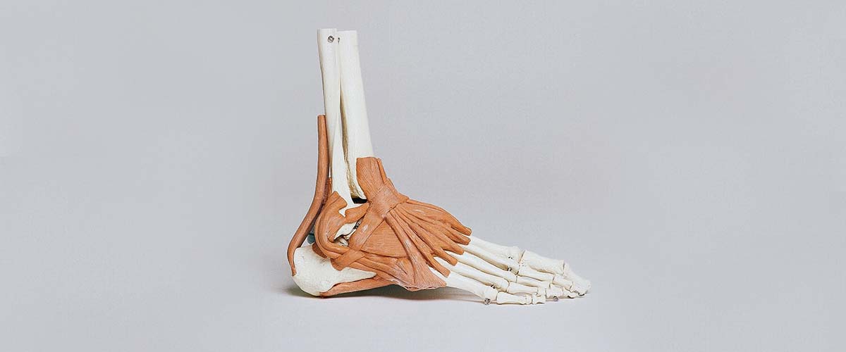
The talus is the bone that links the leg to the foot. Most of it is covered in cartilage, so it has a limited, fragile blood supply. Osteonecrosis (avascular necrosis, AVN) of the talus means part of the bone dies from loss of blood flow. If the dead bone collapses, the ankle and subtalar joints can develop painful arthritis. About 75% of cases follow an injury,1 most often a talus fracture.2
- Deep ankle pain and stiffness months after a talus fracture, or without injury in steroid users
- Osteonecrosis occurred in 31% to 42% after talar neck fractures3,4
- Many cases revascularize without collapse5,6
- Early stages: protected weight-bearing and core decompression12,14
- Collapse: fusion or talus replacement11,16
Causes
- Talus fractures and dislocations: the risk rises with the energy of injury, displacement, comminution and open injuries.1,3 In 798 talar fractures, 42% developed osteonecrosis.3 See foot fractures.
- Atraumatic causes: corticosteroids or high cortisol,9 heavy alcohol use, blood disorders, lupus and other conditions that affect bone blood supply.
Small areas of damaged cartilage and bone on the dome of the talus after a sprain are a different condition, covered on the osteochondral lesions of the talus page.
Symptoms
- Deep, aching ankle pain, worse with walking
- Stiffness and swelling of the ankle
- Pain that persists or returns months after a talus fracture seemed to heal
- With collapse: grinding, deformity and arthritis pain
Diagnosis and the Hawkins sign
After a talus fracture, Dr. Dold watches X-rays for the Hawkins sign: a thin dark line under the dome of the talus about 6 to 9 weeks after injury, which shows the bone still has blood flow. A positive Hawkins sign makes later osteonecrosis unlikely,7 although a few smokers developed it despite the sign.8 MRI is the preferred test for early detection,9 and CT shows collapse and bone loss. Importantly, many talar osteonecroses after fracture revascularize without collapsing: in one series 49% showed osteonecrosis, but more than a third of those recovered.5,6
Treatment before collapse
Evidence favoring one treatment is limited,10,11 but the general approach is:
- Protected weight-bearing in a boot or brace while the bone revascularizes, possibly with shockwave therapy.12
- Core decompression: drilling the dead bone to relieve pressure and stimulate healing.12,13 In one series, 82% of ankles had excellent or good results at about 7 years.14
- Vascularized bone grafting: core decompression with a graft that brings its own blood supply spared more than 80% of patients further surgery in a small series.15
Treatment after collapse
- Fusion: ankle (tibiotalar) or tibiotalocalcaneal fusion is the established salvage operation, relieving pain at the cost of motion.11,12
- Total talus replacement: a custom (often 3D-printed) implant that replaces the whole talus. It is a promising alternative to fusion, improving function scores from 43.2 to 89.3 in pooled studies,16,17 with quick pain relief and preserved motion and limb length.19,20 It can be combined with an ankle replacement.18 Long-term data are still limited.16,18 If you may be a candidate, Dr. Dold can refer you to a surgeon who performs total talus replacement.
Recovery
- Protected weight-bearing: often several months while monitoring with imaging.
- After core decompression or grafting: a period of protected weight-bearing.
- After fusion or replacement: several weeks of non-weight-bearing, then progressive rehab; full recovery takes months.
Dr. Dold reviews your plan based on the stage and your health.
Frequently asked questions
What causes osteonecrosis of the talus?
Most cases follow a talus fracture or dislocation that damages the bone’s blood supply. Others are linked to steroids, alcohol or medical conditions affecting bone circulation.
Will my talus collapse?
Not necessarily. Many cases revascularize without collapse, especially after a positive Hawkins sign on X-ray. Protecting the bone early improves the odds.
What is the Hawkins sign?
A thin dark line under the top of the talus on X-ray about 6 to 9 weeks after a fracture. It means blood flow is returning and makes osteonecrosis unlikely.
Can a damaged talus be replaced?
Yes. A custom total talus replacement is an emerging option that preserves motion, and Dr. Dold can refer you if you may be a candidate. Fusion remains the established alternative.
Is fusion the only option once the bone collapses?
Fusion is reliable for pain relief, but talus replacement, sometimes combined with ankle replacement, is an option for selected patients.
Research cited on this page
- Kopp L, Rammelt S. Posttraumatic Avascular Necrosis of the Talus: Prevention, Course, and Treatment Options. Foot Ankle Clin. 2025;30(1):83-110. PubMed
About 75% of talar osteonecrosis follows trauma; risk rises with more energy, displacement, comminution and soft-tissue or vessel injury. - Martin Oliva X, Viladot Voegeli A. Aseptic (avascular) bone necrosis in the foot and ankle. EFORT Open Rev. 2020;5(10):684-690. PubMed
Talar avascular necrosis most often follows talar fractures, because the talus has a fragile blood supply. - Alley MC, Vallier HA, Tornetta P, et al. Identifying Risk Factors for Osteonecrosis After Talar Fracture. J Orthop Trauma. 2024;38(1):25-30. PubMed
In 798 talar fractures, 42% developed osteonecrosis, more often with neck fractures, displacement and open injuries. - Dodd A, Lefaivre KA. Outcomes of Talar Neck Fractures: A Systematic Review and Meta-analysis. J Orthop Trauma. 2015;29(5):210-5. PubMed
The osteonecrosis rate after talar neck fractures was 31%, rising with Hawkins type. - Vallier HA, Nork SE, Barei DP, et al. Talar neck fractures: results and outcomes. J Bone Joint Surg Am. 2004;86(8):1616-24. PubMed
Osteonecrosis was seen in 49% after talar neck fractures, but over a third revascularized without collapse. - Vallier HA. Fractures of the Talus: State of the Art. J Orthop Trauma. 2015;29(9):385-92. PubMed
Many talar osteonecroses after fracture revascularize without collapse of the talar dome. - Tezval M, Dumont C, Stürmer KM. Prognostic reliability of the Hawkins sign in fractures of the talus. J Orthop Trauma. 2007;21(8):538-43. PubMed
A positive Hawkins sign on X-ray (6 to 9 weeks after injury) makes later osteonecrosis unlikely. - Griffin JT, Landy DC, Mechas CA, et al. The Hawkins Sign of the Talus: The Impact of Patient Factors on Prediction Accuracy. J Bone Joint Surg Am. 2024;106(11):958-965. PubMed
A few patients with a positive Hawkins sign still developed osteonecrosis; all were smokers. - Mousavian A, Abdollahi M, Haddadan N. Talus Avascular Necrosis as a Rare Complication of Cushing’s Disease: A Case Report. Cureus. 2024;16(4):e57531. PubMed
Atraumatic talar osteonecrosis can follow high cortisol (Cushing disease); MRI is preferred for early detection. - Zhang H, Fletcher AN, Scott DJ, et al. Avascular Osteonecrosis of the Talus: Current Treatment Strategies. Foot Ankle Int. 2022;43(2):291-302. PubMed
Treatments range from joint-preserving core decompression to fusion, with newer grafting and talus replacement options; evidence is limited. - Gross CE, Sershon RA, Frank JM, et al. Treatment of Osteonecrosis of the Talus. JBJS Rev. 2016;4(7). PubMed
For early-to-moderate talar osteonecrosis, evidence favoring any one treatment is poor; ankle or hindfoot fusion is a suitable salvage. - Gross CE, Haughom B, Chahal J, et al. Treatments for avascular necrosis of the talus: a systematic review. Foot Ankle Spec. 2014;7(5):387-97. PubMed
Early talar osteonecrosis appears well treated with protected weight-bearing, possibly with shockwave therapy; core decompression is an option if that fails, and fusion is reserved for salvage. - Sultan AA, Mont MA. Core Decompression and Bone Grafting for Osteonecrosis of the Talus: A Critical Analysis of the Current Evidence. Foot Ankle Clin. 2019;24(1):107-112. PubMed
Core decompression and bone grafting have been used for talar osteonecrosis with varying results. - Mont MA, Schon LC, Hungerford MW, et al. Avascular necrosis of the talus treated by core decompression. J Bone Joint Surg Br. 1996;78(5):827-30. PubMed
Core decompression before collapse gave excellent or good results in 82% of ankles at about 7 years. - Cody EA, Nunley JA. Vascularized Pedicle Graft for Talar Osteonecrosis. Foot Ankle Clin. 2019;24(1):121-129. PubMed
Core decompression with a vascularized cuboid bone graft spared more than 80% of patients further surgery in a small series. - Jennison T, Dalgleish J, Sharpe I, et al. Total Talus Replacements. Foot Ankle Orthop. 2023;8(1):24730114221151068. PubMed
Total talus replacement is a promising alternative to fusion for talar osteonecrosis, but more studies are needed. - Bischoff A, Stone R, Dao T, et al. Functional Outcomes and Complications Associated With Total Talus Arthroplasty: A Systematic Review. Foot Ankle Spec. 2023;16(3):259-266. PubMed
After total talus replacement, function scores improved from 43.2 to 89.3 and pain decreased. - Anastasio AT, Bagheri K, Johnson L, et al. Outcomes following total ankle total talus replacement: A systematic review. Foot Ankle Surg. 2024;30(3):245-251. PubMed
Combined total ankle and total talus replacement improved short-term function, but long-term data are lacking. - Kubisa MJ, Kubisa MG, Pałka K, et al. Avascular Necrosis of the Talus: Diagnosis, Treatment, and Modern Reconstructive Options. Medicina (Kaunas). 2024;60(10). PubMed
Talus replacement offers quick pain relief and preserves limb length and motion compared with fusion. - Shnol H, LaPorta GA. 3D Printed Total Talar Replacement: A Promising Treatment Option for Advanced Arthritis, Avascular Osteonecrosis, and Osteomyelitis of the Ankle. Clin Podiatr Med Surg. 2018;35(4):403-422. PubMed
3D-printed total talus replacement is an option for advanced arthritis and talar collapse.
Medically reviewed by Andrew P. Dold, MD, FAOA, FAAOS, FACS, FRCSC. Last reviewed October 2026. This page is general information, not medical advice. Your own diagnosis and treatment plan depend on your exam and imaging.