








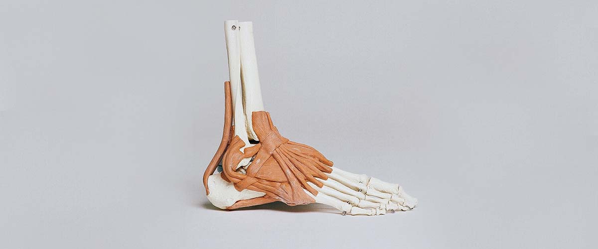
The peroneal tendons run behind the bony bump on the outside of the ankle (the fibula) to the outer foot. They turn the foot outward and help keep the ankle from rolling.1 Peroneal problems range from tendonitis to split tears and tendons that snap out of their groove,2 and they are often mistaken for an ankle sprain, so they can go unrecognized for months.5,6
- Pain and swelling behind and below the outer ankle bone
- Often follows ankle sprains or occurs with a high-arched (varus) heel2,6
- Ultrasound and MRI confirm tears and slipping tendons4,10
- Most tendonitis improves with a brace, lateral-wedge insert and rehab6,7
- Tears that don’t heal and tendons that dislocate usually need surgery15,24
Types of peroneal tendon problems
- Tendonitis and tenosynovitis: irritation of the tendons and their sheath, usually from overuse.7
- Peroneus brevis split tears: a lengthwise tear where the tendon wraps around the tip of the fibula.15
- Peroneus longus tears: often farther down, near the cuboid bone, sometimes with a painful or broken os peroneum (a small bone inside the tendon).12,15
- Subluxation and dislocation: the tendons snap forward over the fibula when the retinaculum (the band that holds them in place) is torn, typically from a forceful push-off or landing,7 or slip within their own sheath.10
Who gets them?
Peroneal problems are common in running, cutting and jumping sports and after repeated ankle sprains. A high-arched foot with a heel that tilts inward (hindfoot varus) and chronic lateral ankle instability both increase the load on the tendons.2,3,6 Persistent pain after ankle ligament surgery is also common, and peroneal tendon problems are one of the causes to check.31
Symptoms
- Pain and swelling behind or below the outer ankle bone
- Pain when pushing off, cutting or walking on uneven ground
- A snapping or popping feeling on the outside of the ankle
- Weakness or a sense the ankle wants to give way
Diagnosis
Dr. Dold tests the tendons for tenderness, strength and stability, checks for snapping as you circle your ankle, and looks at the shape of your foot and the stability of your ankle ligaments. Imaging helps:
- Ultrasound, especially dynamic ultrasound while you move the ankle, can show tendons slipping out of position.10,11
- MRI shows tears, tenosynovitis and related problems such as ligament or cartilage injuries.4 Specific MRI sequences help avoid an artifact that can make normal tendon look abnormal.9
- X-rays look for a fractured os peroneum, which can signal a peroneus longus tear,12 and for heel and foot alignment.
Non-surgical treatment
Most peroneal tendonitis is treated without surgery first:6,7
- Rest from aggravating activity, sometimes with a walking boot for a short period
- An ankle brace and a lateral-wedge insert to unload the tendons, especially with a varus heel
- Physical therapy with progressive tendon loading, balance and strengthening
- Anti-inflammatory medicines when safe for you
A cortisone injection into the tendon sheath can help selected patients: in one series, 36.8% had more than 12 weeks of relief and complications were rare, though 25% later had surgery.14
Surgery for peroneal tendon tears
When pain continues despite good non-surgical care, or a tear is significant, surgery is considered. Most acute peroneus brevis tears are longitudinal and often need repair.15 Options include:
- Debridement and repair (tubularization) for less extensive tears.16
- Tenodesis (sewing the damaged tendon to the healthy one) for severe tears.16
- Reconstruction or tendon transfer when the tendon cannot be repaired.22
- Peroneal tendoscopy (a small camera in the tendon sheath) for synovitis, scar tissue and some tears, with meaningful functional gains and few adverse events.29,30
Across studies, scores improved substantially after surgery,17 and most patients report high satisfaction and return to their prior function.18 Peroneus longus repairs have good outcomes with strength similar to the other foot,19,20 and acute tears tend to do better than long-standing ones.21 Underlying problems such as a varus heel or ankle instability are corrected at the same time when present.2,3
Peroneal tendon subluxation and dislocation
Tendons that snap out of the groove behind the fibula often don’t settle with non-surgical care. Non-surgical treatment of acute dislocation has failure rates up to 50%,24 and with taping the tendons redislocated in 60%; a non-weight-bearing cast for 6 weeks or more worked better.23 For recurrent or chronic instability, surgery repairs the retinaculum, often with deepening of the groove behind the fibula. Redislocation after surgery is under 1.5%, and combining groove deepening with retinaculum repair was linked to higher return to sport.25 Studies report excellent or good results in about 90% of athletes,26 and tendoscopic (minimally invasive) retinaculum repair is an option in selected cases.28
Recovery
- Tendonitis: most people improve over weeks to a few months with bracing and rehab.
- After tear repair or stabilization: a splint and boot with protected weight-bearing for several weeks, then physical therapy for motion, strength and balance.
- Return to sport after surgery usually takes a few months and depends on the procedure.
Dr. Dold reviews your expected timeline based on your injury and treatment.
Frequently asked questions
Is peroneal tendonitis the same as an ankle sprain?
No, but they often happen together and are easy to confuse. Pain that lingers behind the outer ankle bone after a sprain may come from the peroneal tendons.
Can a peroneal tendon tear heal without surgery?
Mild tendinopathy and small tears often improve with bracing, inserts and rehab. Larger or painful split tears that don’t respond usually need surgery.
Why does my ankle snap on the outside?
A snapping feeling can mean the peroneal tendons are slipping out of their groove behind the fibula, often after a torn retinaculum. Dynamic ultrasound can show it.
Does a high arch matter?
Yes. A high-arched foot with an inward-tilted heel loads the peroneal tendons and is a known risk factor. Inserts help, and the alignment may be corrected at surgery.
How long is recovery after peroneal tendon surgery?
Usually several weeks of protected weight-bearing followed by therapy, with return to sport after a few months depending on the procedure.
Research cited on this page
- Philbin TM, Landis GS, Smith B. Peroneal tendon injuries. J Am Acad Orthop Surg. 2009;17(5):306-17. PubMed
The peroneal tendons are the main evertors of the foot and help stabilize the outside of the ankle. - Roster B, Michelier P, Giza E. Peroneal Tendon Disorders. Clin Sports Med. 2015;34(4):625-41. PubMed
Peroneal problems include tendinosis, tenosynovitis, tears, subluxation and painful os peroneum; hindfoot varus and ankle instability predispose. - Zgonis T, Jolly GP, Polyzois V, et al. Peroneal tendon pathology. Clin Podiatr Med Surg. 2005;22(1):79-85. PubMed
Chronic lateral ankle instability and excess varus can damage the peroneal tendons. - Davda K, Malhotra K, O’Donnell P, et al. Peroneal tendon disorders. EFORT Open Rev. 2017;2(6):281-292. PubMed
Ultrasound and MRI help confirm the diagnosis; treatment recommendations rest mainly on case series and opinion. - Mostovoy A, Chang T. Peroneal Pathology in the Athlete. Clin Podiatr Med Surg. 2023;40(1):139-155. PubMed
Peroneal problems in athletes are commonly misdiagnosed and can cause chronic pain. - Deu RS, Coslick AM, Dreher G. Tendinopathies of the Foot and Ankle. Am Fam Physician. 2022;105(5):479-486. PubMed
Peroneal problems are often mistaken for ankle sprains; varus hindfoot is a risk factor; treatment includes immobilization, laterally posted orthotics and progressive tendon loading. - Lugo-Pico JG, Kaiser JT, Sanchez RA, et al. Peroneal Tendinosis and Subluxation. Clin Sports Med. 2020;39(4):845-858. PubMed
Tendinopathy usually comes from overuse; conservative care is first-line, while subluxation often needs surgery in athletes. - van Dijk PA, Madirolas FX, Carrera A, et al. Peroneal tendons well vascularized: results from a cadaveric study. Knee Surg Sports Traumatol Arthrosc. 2016;24(4):1140-7. PubMed
In a cadaver study the peroneal tendons were well vascularized overall, though 8 of 10 longus tendons had poorly vascularized zones. - Ieong E, Rafferty M, Khanna M, et al. Use of Fat-Suppressed T2-Weighted MRI Images to Reduce the Magic Angle Effect in Peroneal Tendons. Foot Ankle Spec. 2019;12(6):513-517. PubMed
Adding T2-weighted MRI sequences reduced a common MRI artifact (magic angle) in the peroneal tendons. - Draghi F, Bortolotto C, Draghi AG, et al. Intrasheath Instability of the Peroneal Tendons: Dynamic Ultrasound Imaging. J Ultrasound Med. 2018;37(12):2753-2758. PubMed
Dynamic ultrasound is the imaging modality of choice for tendons that slip within their sheath. - Pesquer L, Guillo S, Poussange N, et al. Dynamic ultrasound of peroneal tendon instability. Br J Radiol. 2016;89(1063):20150958. PubMed
Dynamic ultrasound allows real-time diagnosis and classification of peroneal tendon instability. - Brigido MK, Fessell DP, Jacobson JA, et al. Radiography and US of os peroneum fractures and associated peroneal tendon injuries: initial experience. Radiology. 2005;237(1):235-41. PubMed
Wide separation of a fractured os peroneum on X-ray was associated with a full-thickness peroneus longus tear in all 7 cases. - Mizel MS, Michelson JD, Newberg A. Peroneal tendon bupivacaine injection: utility of concomitant injection of contrast material. Foot Ankle Int. 1996;17(9):566-8. PubMed
Contrast injected with a diagnostic numbing shot leaked into nearby joints in 15%, so the test may not be 100% accurate. - Fram BR, Rogero R, Fuchs D, et al. Clinical Outcomes and Complications of Peroneal Tendon Sheath Ultrasound-Guided Corticosteroid Injection. Foot Ankle Int. 2019;40(8):888-894. PubMed
After a peroneal sheath injection, 36.8% had more than 12 weeks of relief, 25% later had surgery, and complications were rare. - Slater HK. Acute peroneal tendon tears. Foot Ankle Clin. 2007;12(4):659-74, vii. PubMed
Most acute peroneus brevis tears are longitudinal near the tip of the fibula and often need surgery; longus tears may first be managed without surgery. - Dombek MF, Orsini R, Mendicino RW, et al. Peroneus brevis tendon tears. Clin Podiatr Med Surg. 2001;18(3):409-27. PubMed
For less extensive brevis tears, debridement and tubularization are recommended; severe tears may need tenodesis. - Mercer NP, Gianakos AL, Mercurio AM, et al. Clinical Outcomes of Peroneal Tendon Tears: A Systematic Review. J Foot Ankle Surg. 2021;60(5):1008-1013. PubMed
After surgery for peroneal tears, scores improved from 41.1 to 84.4, with an overall complication rate of 38.7%, mostly minor. - Bahad SR, Kane JM. Peroneal Tendon Pathology: Treatment and Reconstruction of Peroneal Tears and Instability. Orthop Clin North Am. 2020;51(1):121-130. PubMed
Patients report high satisfaction after surgery, and most return to their pre-injury function. - Burkhard MD, Wirth SH, Andronic O, et al. Clinical and Functional Outcomes of Peroneus Longus to Brevis Tendon Transfer. Foot Ankle Int. 2021;42(6):699-705. PubMed
After peroneus longus surgery, outcome scores were good and strength matched the other foot. - Arbab D, Tingart M, Frank D, et al. Treatment of isolated peroneus longus tears and a review of the literature. Foot Ankle Spec. 2014;7(2):113-8. PubMed
Isolated peroneus longus tears were diagnosed an average of 11 months after injury; 5 of 6 had good or excellent results after surgery. - Sammarco GJ. Peroneus longus tendon tears: acute and chronic. Foot Ankle Int. 1995;16(5):245-53. PubMed
Patients with acute peroneus longus tears tended to do better than those with chronic tears. - Sherman TI, Koury K, Orapin J, et al. Lateral Transfer of the Flexor Digitorum Longus for Peroneal Tendinopathy. Foot Ankle Int. 2019;40(9):1012-1017. PubMed
Transfer of the flexor digitorum longus tendon was an effective option for irreparable peroneal tendons in a small series. - Bakker D, Schulte JB, Meuffels DE, et al. Non-operative treatment of peroneal tendon dislocations: A systematic review. J Orthop. 2020;18:255-260. PubMed
Without surgery, tendon dislocation recurred in 60% treated with taping; a non-weight-bearing cast for 6 or more weeks worked in 5 of 6. - Espinosa N, Maurer MA. Peroneal tendon dislocation. Eur J Trauma Emerg Surg. 2015;41(6):631-7. PubMed
Non-surgical treatment of acute peroneal dislocation has failure rates up to 50%; chronic instability usually needs surgery. - van Dijk PA, Gianakos AL, Kerkhoffs GM, et al. Return to sports and clinical outcomes in patients treated for peroneal tendon dislocation: a systematic review. Knee Surg Sports Traumatol Arthrosc. 2016;24(4):1155-64. PubMed
After surgery for peroneal dislocation, redislocation was under 1.5%; groove deepening plus retinaculum repair had higher return to sport. - Guillo S, Calder JD. Treatment of recurring peroneal tendon subluxation in athletes: endoscopic repair of the retinaculum. Foot Ankle Clin. 2013;18(2):293-300. PubMed
Surgery for recurring peroneal subluxation in athletes reported excellent or good results in about 90%. - Ferran NA, Oliva F, Maffulli N. Recurrent subluxation of the peroneal tendons. Sports Med. 2006;36(10):839-46. PubMed
Small series cannot show which procedure for recurrent peroneal subluxation works better; anatomic retinaculum repair seems appropriate. - Nishimura A, Kato K, Nakazora S, et al. Tendoscopic peroneal retinaculum repair for recurrent peroneal tendon dislocation enables earlier return to sports than the open procedure. Knee Surg Sports Traumatol Arthrosc. 2020;28(10):3318-3323. PubMed
Tendoscopic retinaculum repair for recurrent dislocation was less invasive and allowed an early return to sports. - Tham A, Rajivan R, Rubin J, et al. Peroneal tendoscopy for peroneal tendon disorders: A systematic review of indications, diagnostic utility, and clinical outcomes. J Orthop. 2026;74:24-32. PubMed
Peroneal tendoscopy gave meaningful functional gains, timely return to sport and few adverse events. - Lui TH, Tse LF. Peroneal tendoscopy. Foot Ankle Clin. 2015;20(1):15-25. PubMed
Tendoscopy is used for tenosynovitis, subluxation, snapping, partial tears and scarring, with good to excellent outcomes in most. - Ahn BH, Cho BK. Persistent Pain After Operative Treatment for Chronic Lateral Ankle Instability. Orthop Res Rev. 2021;13:47-56. PubMed
Persistent pain after lateral ankle ligament surgery is common (13% to 35%), and peroneal problems are one cause.
Medically reviewed by Andrew P. Dold, MD, FAOA, FAAOS, FACS, FRCSC. Last reviewed October 2026. This page is general information, not medical advice. Your own diagnosis and treatment plan depend on your exam and imaging.